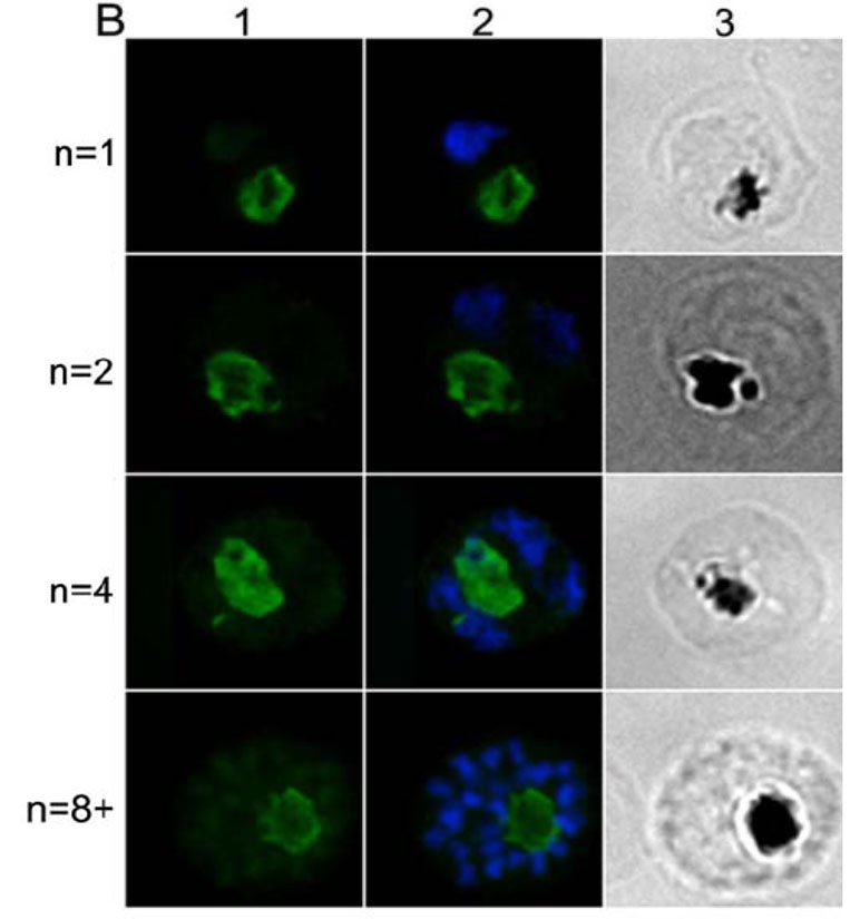
IFA location of CRT in mature parasites at different stages of nuclear division. Each row in panel B shows an identical field from 1% formaldehyde-fixed thin smears of late-stages parasites at the 1-, 2-, 4-, and 8-nuclei stage. Panel B1 shows rabbit anti-CRT antibody labeling (the secondary antibody was Alexa Fluor 488-conjugated anti-rabbit IgG antibody (green). Panel B2 shows the same fields merged with the corresponding DAPI stained nucleus images (blue). Panel B3 shows the same fields visualized by light microscopy - note the pigment is clearly visible. Rabbit-anti-CRT antiobody is clearly detectable throughout parasite maturation and is associated with the food vacuole.
Dluzewski AR, Ling IT, Hopkins JM, Grainger M, Margos G, Mitchell GH, Holder AA, Bannister LH. Formation of the food vacuole in Plasmodium falciparum: a potential role for the 19 kDa fragment of merozoite surface protein 1 (MSP1(19)). PLoS ONE. 2008 Aug 3(8):e3085. ™
