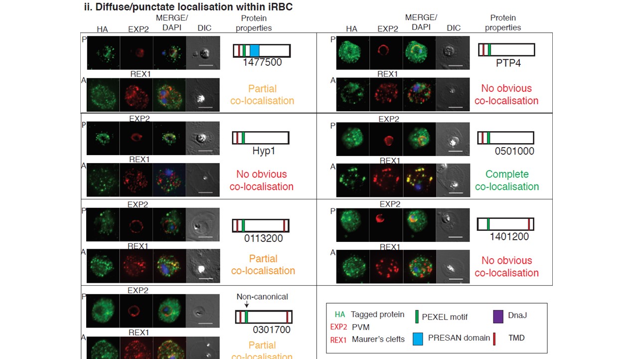
Illumination Microscopy (SIM) analysis was used to obtain higher spatial resolution. Seven proteins showed diffuse iRBC signal resembling Maurer’s clefts and showed either no, partial or complete co-localisation with the MCs marker REX1. Scale bars= 5 µm or 1 µm
Jonsdottir TK, Counihan NA, Modak JK, Kouskousis B, Sanders PR, Gabriela M, Bullen HE, Crabb BS, de Koning-Ward TF, Gilson PR. Characterisation of complexes formed by parasite proteins exported into the host cell compartment of Plasmodium falciparum infected red blood cells. Cell Microbiol. 2021 28:e13332.
Other associated proteins
| PFID | Formal Annotation |
|---|---|
| PF3D7_0113200 | Plasmodium exported protein, unknown function |
| PF3D7_0113300 | Plasmodium exported protein (hyp1), unknown function |
| PF3D7_0301700 | Plasmodium exported protein, unknown function |
| PF3D7_0501000 | Plasmodium exported protein, unknown function |
| PF3D7_0935900 | ring-exported protein 1 |
| PF3D7_1401200 | Plasmodium exported protein, unknown function |
| PF3D7_1471100 | exported protein 2 |
| PF3D7_1477500 | Plasmodium exported protein (PHISTb), unknown function |
