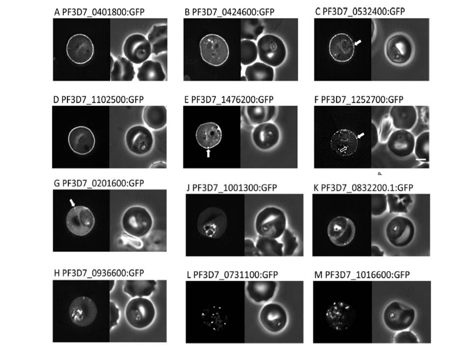
Localisation of PHIST:GFP proteins. The left- and right-hand images show GFP localisation and a phase contrast image, respectively. White filled arrows: peripheral GFP puncta; White unfilled arrow: GFP puncta in host erythocyte cytosol. Scale bar, 2 mm. GFP-tagged PF3D7_0401800, PF3D7_0424600, PF3D7_0532400, PF3D7_1102500 and PF3D7_1476200 were all exported to the host cell and displayed a striking localisation at the edge of the host erythrocyte (1A–E), PF3D7 1252700 was clearly peripheral in the host erythrocyte (F). PF3D7_0201600 (G) shows weak accumulation at the erythrocyte periphery, but the majority of the protein was localised in the RBC cytosol. PF3D7_0936600 was localised in the erythrocyte cytosol, not at the host cell periphery (H).
Tarr SJ, Moon RW, Hardege I, Osborne AR. A conserved domain targets exported PHISTb family proteins to the periphery of Plasmodium infected erythrocytes. Mol Biochem Parasitol. 2014 196(1):29-40 PMID: 25106850
Other associated proteins
| PFID | Formal Annotation |
|---|---|
| PF3D7_0201600 | PHISTb domain-containing RESA-like protein 1 |
| PF3D7_0401800 | Plasmodium exported protein (PHISTb), unknown function |
| PF3D7_0424600 | plasmodium exported protein (phistb) |
| PF3D7_0532400 | lysine-rich membrane-associated PHISTb protein |
| PF3D7_0832200 | Plasmodium exported protein (PHISTa-like), unknown function |
| PF3D7_0936600 | Plasmodium exported protein (PHISTc), unknown function gametocyte exported protein 5 |
| PF3D7_1001300 | Plasmodium exported protein (PHISTa), unknown function |
| PF3D7_1016600 | Plasmodium exported protein (PHISTc), unknown function |
| PF3D7_1102500 | gametocyte exported protein 2 Plasmodium exported protein (PHISTb), unknown function |
| PF3D7_1252700 | Plasmodium exported protein (PHISTb), unknown function |
| PF3D7_1476200 | Plasmodium exported protein (PHISTb), unknown function |
