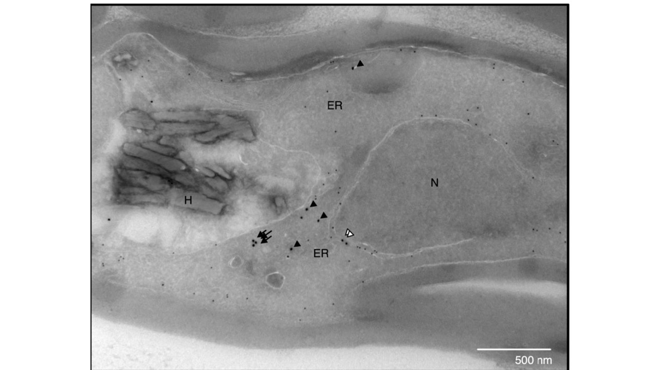
Localisation of AP-2μ-2xFKBPP-GFP and PDI by immunoelectron microscopy. Electron micrographs of vesicular colocalisation of AP-2μ-2xFKBP-GFP (18 nm gold particles) and PDI (12 nm gold particles) in developing trophozoites. N: nucleus; ER: endoplasmic reticulum; H: haemozoin; white arrowhead: AP-2μ at the nuclear membrane; black arrow: AP-2μ in vesicles; black arrowhead: AP-2μ at the ER. Immunostaining against the endoplasmic reticulum (ER)-resident protein disulfide isomerase (PDI) suggests that these tubular structures may be cross-sections of distal extensions of the ER. Parasites expressing AP-2µ-2xFKBP-GFP showed a similar localisation by immuno-EM but GFP fluorescence was too faint to reliably observe in live cells.
Modification of an atypical clathrin-independent AP-2 adaptin complex of Plasmodium falciparum reduces susceptibility to artemisinin. Ryan C. Henrici, Rachel L. Edwards, Martin Zoltner, Donelly A. van Schalkwyk, Melissa N. Hart, Franziska Mohring, Robert W. Moon, Stephanie D. Nofal, Avnish Patel, Christian Flueck, David A. Baker
Other associated proteins
| PFID | Formal Annotation |
|---|---|
| PF3D7_1218300 | AP-2 complex subunit mu |
