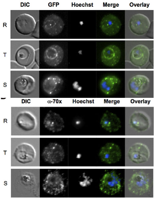
PfHsp70-x localizes to the host cell and PV. Upper panel: Live cell imaging of 3D7G:70x-infected erythrocytes. In the merge and overlay images: green, GFP; blue, Hoechst. In all stages punctate GFP fluorescence can be visualized in the host cell. Additionally, a ring of fluorescence indicative of a PV localization can also be seen. Lower panel: Indirect immunofluorescence localization of PfHsp70-x. In the merge and overlay images: green, a-70x/MRax; blue, Hoechst. R, ring stage; T, trophozoite stage; S, schizont stage.
Külzer S, Charnaud S, Dagan T, Riedel J, Mandal P, Pesce ER, Blatch GL, Crabb BS, Gilson PR, Przyborski JM. Plasmodium falciparum-encoded exported hsp70/hsp40 chaperone/co-chaperone complexes within the host erythrocyte. Cell Microbiol. 2012 14(11):1784-95
