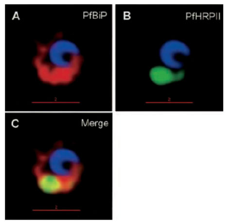
Protein accumulation in the ER in P. falciparum-infected cells treated with brefeldin A. Infected red cells at early times of asexual growth, were incubated with 5 mgml-1 brefeldin A for 24 h and subsequently stained for PfHRPII (green), PfBiP (red), Hoechst (blue for nuclei), in indirect immunofluorescence assays and imaged by DeltaVision deconvolution microscopy. Yellow indicates extent of merge between green and red. The location to the ER may reflect altered (albeit reversible) membrane organization induced by the drug, rather than a specialized organelle. In addition, since the P. falciparum (primary) ER is not well characterized, the context of a secondary ER remains unclear.
Haldar K, Mohandas N, Samuel BU, Harrison T, Hiller NL, Akompong T, Cheresh P. Protein and lipid trafficking induced in erythrocytes infected by malaria parasites. Cell Microbiol. 2002 4:383-95.
Other associated proteins
| PFID | Formal Annotation |
|---|---|
| PF3D7_1108600 | endoplasmic reticulum-resident calcium binding protein |
