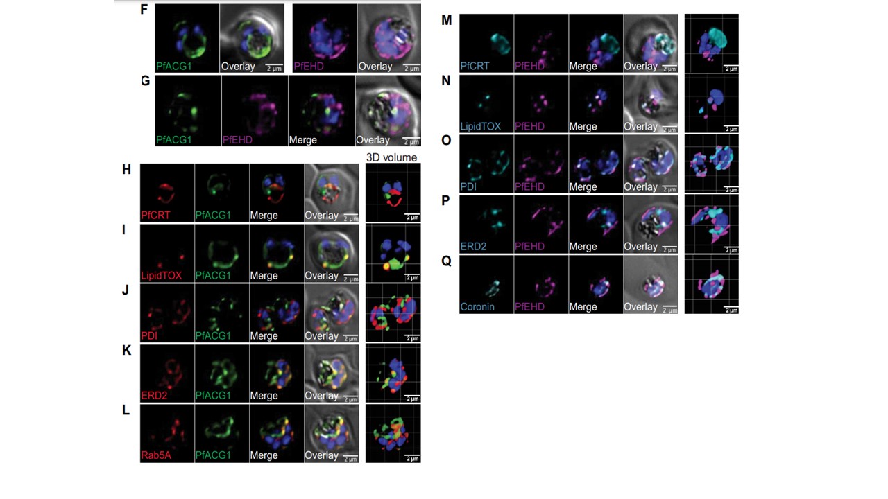
(F) Fluorescence microscopy images of fixed NF543×HA-EHDattB-ACG1-eGFP parasites stained with either antiGFP (green) antibodies or anti-HA (magenta) antibodies. Nuclei were stained with DAPI (blue). Scale bars, 2 mm. (G) Fluorescence microscopy image of fixed and doubly stained NF543×HA-EHDattB-ACG1-eGFP parasites using anti-GFP (green) and anti-HA (magenta) antibodies. Nuclei were stained with DAPI (blue). Scale bar, 2 mm. (H to L) Fluorescence microscopy images and 3D reconstructions of fixed NF543×HA-EHDattB-ACG1-eGFP parasites costained with antibodies to anti-GFP (green) and (H) anti-PfCRT antibodies, (I) LipidTOX neutral lipid stain, (J) anti-PDI, (K) anti-ERD2, or (L) anti-Rab5A (red) antibodies. Nuclei were stained with DAPI (blue). Scale bars, 2 mm. (M to Q) Fluorescence microscopy images and 3D reconstructions of fixed NF543×HA-EHDattB-ACG1-eGFP parasites costained with antibodies to anti-HA (magenta) and (M) anti-PfCRT antibodies, (N) LipidTOX neutral lipid stain, (O) anti-PDI, (P) anti-ERD2, or (Q) anti-coronin (cyan) antibodies. Nuclei were stained with DAPI (blue). Scale bars, 2 mm.
Murithi JM et al The antimalarial MMV688533 provides potential for single-dose cures with a high barrier to Plasmodium falciparum parasite resistance. Sci Transl Med. 2021 13(603):eabg6013
Other associated proteins
| PFID | Formal Annotation |
|---|---|
| PF3D7_0211200 | ras-related protein Rab-5A |
| PF3D7_0304200 | EH domain-containing protein |
| PF3D7_0709000 | chloroquine resistance transporter |
| PF3D7_0827900 | protein disulfide isomerase |
| PF3D7_1251200 | coronin |
| PF3D7_1353600 | ER lumen protein retaining receptor |