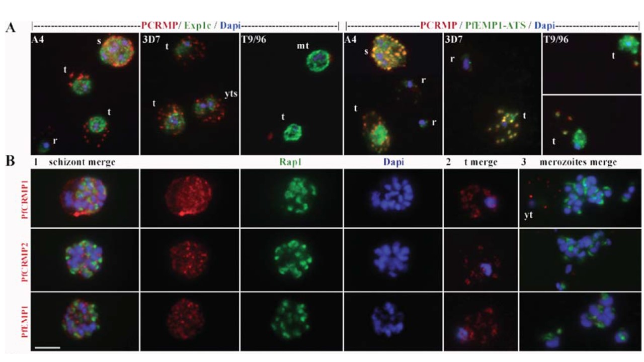
PfCRMP1 and 2 are expressed in blood-stage parasites and colocalize with PfEMP1. A. Merged images of the fluorescence patterns of antisera detecting PCRMP1 (red) and the PVM antigen, EXP1C, or PfEMP1-ATS (green) as indicated show that PCRMP1 is exported to structures within P. falciparum A4-, 3D7- and T9-96-IE cytoplasm and colocalizes with PfEMP1 in the MCs of IEs. Nuclei are stained with DAPI (blue). PfCRMP1 and PfEMP1 colocalize in abnormal aggregates in the cytoplasm of T9/96 mature trophozoite-IEs. B. PfCRMP1, PfCRMP2 and PfEMP1 are detected in an indistinguishable, diffuse, punctate pattern at the surface of P. falciparum A4 schizonts (1) and in the MCs of young trophozoite-IEs (2) but are not associated with released merozoites (3). Merged and individual images of the florescence pattern of parasites stained with the anti-peptide antibodies (red) indicated on the LHS and antiserum detecting the rhoptry protein RAP1 (green). r, rings; yt, young trophozoites; t, trophozoite; mt, mature trophozoites; s, schizonts. Scale bar = 5 mm.
Kafatos FC, Janse CJ, Waters AP. Plasmodium cysteine repeat modular proteins 1-4: complex proteins with roles throughout the malaria parasite life cycle. Cell Microbiol. 2007 9(6):1466-80.
Other associated proteins
| PFID | Formal Annotation |
|---|---|
| PF3D7_0718300 | cysteine repeat modular protein 2 |
| PF3D7_1410400 | rhoptry-associated protein 1 |
| PfEMP1 | PfEMP1 |
