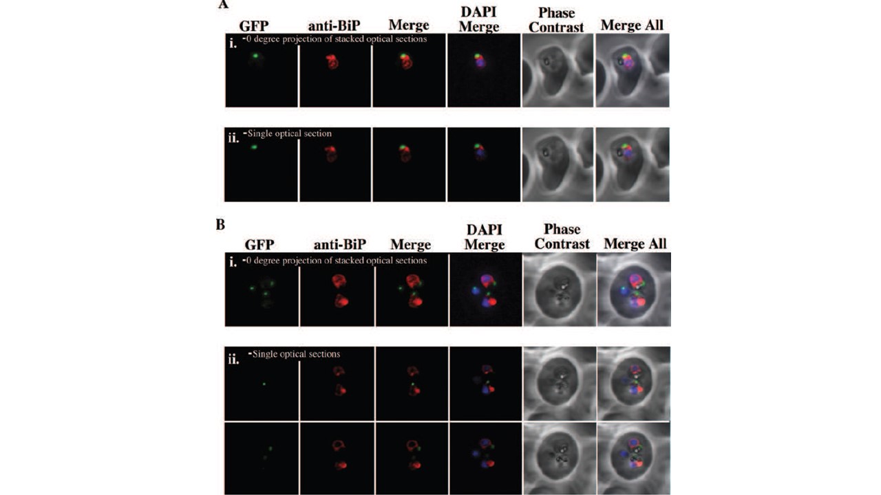
The apicoplast has a close association with the ER. ACP(l)-GFP expressing parasites (GFP in the apicoplast) were analysed by IFA with anti-BiP antibody labelling to assess the relationship between the ER and the apicoplast. A. Anti-BiP IFA of a ring-stage parasite expressing ACP(l)-GFP. (i) A 0° projection of stacked optical sections demonstrating that the apicoplast
is ‘cupped’ by a strand of ER. (ii) A single optical section demonstrating that the apicoplast and ER are closely juxtaposed. A 360° rotation of this sample can be viewed in B. Anti-BiP IFA of multiple ACP(l)-GFP expressing parasites within the same erythrocyte. (i) A 0° projection of stacked optical sections showing that each apicoplast is closely associated with a strand of ER. (ii) Two single optical sections focus on each parasites apicoplast and show that the apicoplast and the ER are closely apposed. The third parasite in the erythrocyte is devoid of ER and is probably dead. Tonkin CJ, Struck NS, Mullin KA, Stimmler LM, McFadden GI. Evidence for Golgi-independent transport from the early secretory pathway to the plastid in malaria parasites. Mol Microbiol. 2006 61(3):614-30. PMID: 16787449.
