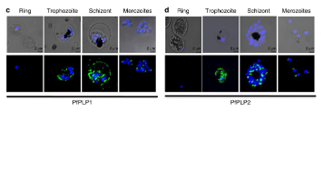
Schematic representation of all PfPLPs (PfPLP1 (PFD0430c), PfPLP2 (PFL0805w), PfPLP3 (PFI1145w), PfPLP4 (Pf08_0050) and PfPLP5 (Pf08_0052)). The signal sequence (dark grey box) and MACPF (Membrane attack complex/Perforin) domain (Light grey box) are represented. The MACPF domain includes a conserved signature motif (shown in yellow box), and two transmembrane helical domains, CH1 and CH2 (shown in red boxes). The region underlined denotes the region of the protein against which antibodies have been raised. Scale bar, 100 aa. (b) Expression of PfPLPs in the blood stage of P. falciparum. P. falciparum culture containing rings, trophozoites and schizonts (iRBC) was saponin treated, lysed and probed with antisera of different PfPLPs by western blotting. PfPLP1 and PfPLP2 were detected in the western blot corresponding to the full-length size of ~95 and ~125 kD respectively. RBC lysate was also probed with antisera of PfPLP’s as a negative control. (c,d) Stage-specific expression of PfPLP1 and PfPLP2 in the blood stage of P. falciparum. Expression of PfPLP1 and PfPLP2 starts in trophozoite stage, with maximal expression in schizonts. In mature schizonts, PfPLP1 and PfPLP2 show punctate staining at the apical end of merozoites. PfPLP1 also shows a ring-like staining, suggesting its localization to RBCM. Scale bar, 2 μm. Garg S, Agarwal S, Kumar S, Yazdani SS, Chitnis CE, Singh S. Calcium-dependent permeabilization of erythrocytes by a perforin-like protein during egress of malaria parasites. Nat Commun. 2013
Other associated proteins
| PFID | Formal Annotation |
|---|---|
| PF3D7_0408700 | perforin-like protein 1 |
| PF3D7_0819400 | perforin-like protein 4 |
| PF3D7_1216700 | perforin-like protein 2 |
