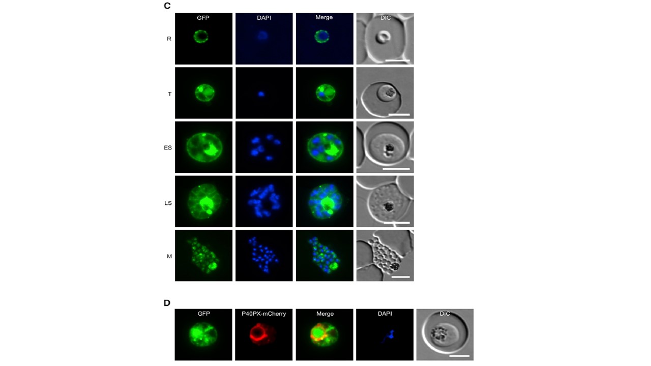
Endogenous Tagging and Localization Analysis of PfLCN (lipocalin). Integration PCR of transgenic LCN-Kd and LCN-Con knockin (KI) and unmodified wild-type (WT) parasites. (C and D) Localization of PfLCN-GFP (green) by live-cell microscopy. In (C), the localization in ring (R), trophozoite (T), early schizont (ES), and late schizont (LS) stage parasites as well as in free merozoite (M) is shown. In (D), the FV membrane marker P40PX-mCherry (red) was coexpressed in LCN-Kd parasites. Nuclei were stained with DAPI (blue). DIC, differential interference contrast. Scale bar, 5 mm. Live-cell microscopy of LCN-Kd parasites revealed that in rings and early trophozoites PfLCN-GFP localized to the parasite periphery, suggesting a possible localization on the PVM, the PPM, or within the PV itself (C). In trophozoites and early schizonts, the protein was additionally visible in small, mostly peripheral, focal structures, in accumulations in the parasite cytoplasm as well as in the FV. The localization to the FV in trophozoites was further verified using the FV membrane marker P40PX-mCherry, which was co-expressed in LCN-Kd parasites (D). In schizonts, we mainly observed the PfLCN signal around developing merozoites, in some accumulations in the schizont cytoplasm and in the FV. In contrast, in free merozoites PfLCN was present in the cytosol but no GFP signal was evident around their periphery, excluding a localization to the PPM and further supporting that PfLCN localizes to the PV and possibly on the PVM of the infected erythrocyte (C).
Burda PC, Crosskey T, Lauk K, Zurborg A, Söhnchen C, Liffner B, Wilcke L,Pietsch E, Strauss J, Jeffries CM, Svergun DI, Wilson DW, Wilmanns M, Gilberger TW. Structure-Based Identification and Functional Characterization of a Lipocalin in the Malaria Parasite Plasmodium falciparum. Cell Rep. 2020 31(12):107817.