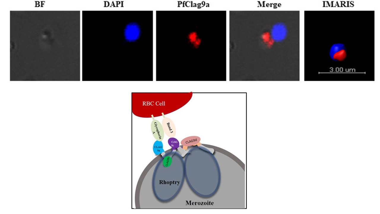
Up: Expression of PfClag9 gene fragments and localization of PfClag9a on merozoite surface. Indirect immunofluorescence assay (IFA) and confocal microscopy of P. falciparum-infected merozoites. P. falciparum parasites were probed using mice anti-PfClag9a antibody followed by FITC-conjugated using secondary anti-mice IgG’s. PfRhopH3 was used as a marker for rhoptry protein. The parasite nuclei were counter stained with DAPI (blue) and slides were visualized by confocal laser scanning microscope. As a negative control, pre-immune antibodies were used to stain the fixed infected RBCs. To determine the cellular localization of PfClag9, intact merozoites were stained with each of the four PfClag9 antibodies raised in mice. All four anti-mice PfClag9 antibody preparations stained merozoites at the apical end of the parasites. Righ: A model of PfRhopH-Clag9 complex based on protein- protein interactions and protein-erythrocyte interaction
Chourasia BK, Deshmukh A, Kaur I, Paul G, Panda A, Rathore S, Singh SK, Theisen M, Mohmmed A, Malhotra P. Plasmodium falciparum Clag9 associated PfRhopH complex is involved in merozoite binding to human erythrocytes. Infect Immun. 2019 Nov 11. pii: IAI.00504-19.
