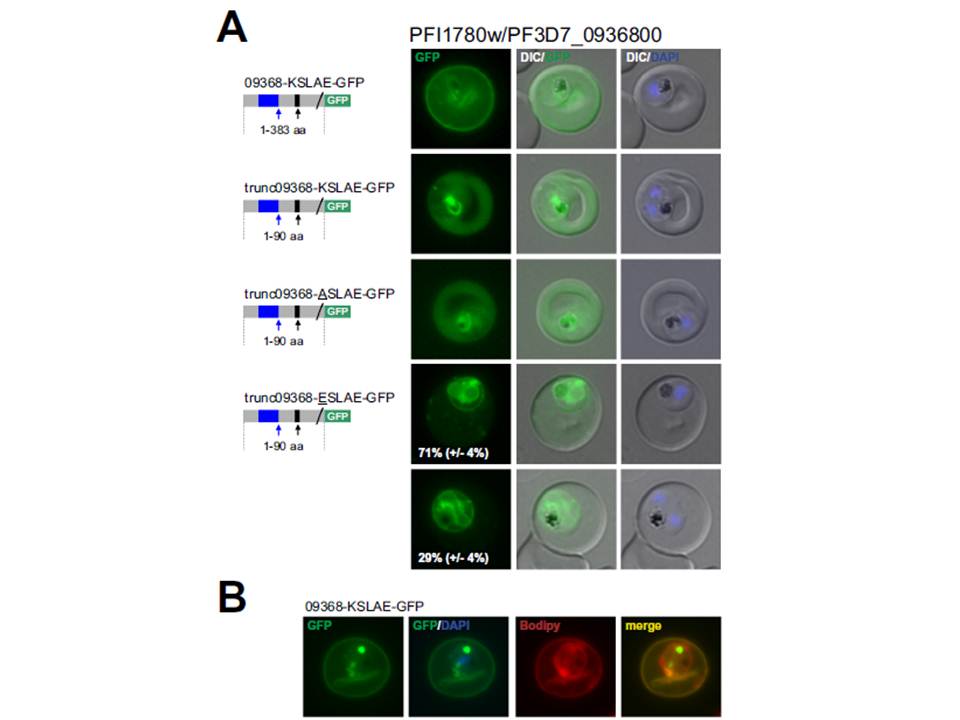
PFI1780w/PF3D7_0936800 encodes an exported PHISTc protein that is cleaved at the non-canonical PEXEL/HT. A. Fluorescence microscopy. The schematic structure of the GFP-tagged proteins is depicted on the left of representative images of iRBCs. The ER-signal is shaded blue, the (non-) canonical PEXEL/HT is shaded black and GFP is shaded green. Putative cleavage sites are indicated by an arrow (blue: SP cleavage site; black: Plasmepsin V cleavage site. For trunc09368-ESLAE-GFP two panels are shown representing iRBCs with reduced export and no export of the reporter protein (at least 50 iRBCs were analyzed on four occasions, whereas ratio is indicated in %; standard deviation in brackets). B. Bodipy-TR-C5-ceramid stained iRBCs expressing 09368_KSLAE-GFP. Fluorescence was located close to the RBC membrane
Schulze J, Kwiatkowski M, Borner J, Schlüter H, Bruchhaus I, Burmester T, Spielmann T, Pick C. The Plasmodium falciparum exportome contains non-canonical PEXEL/HT proteins. Mol Microbiol. 2015 Apr 8. [Epub ahead of print]
