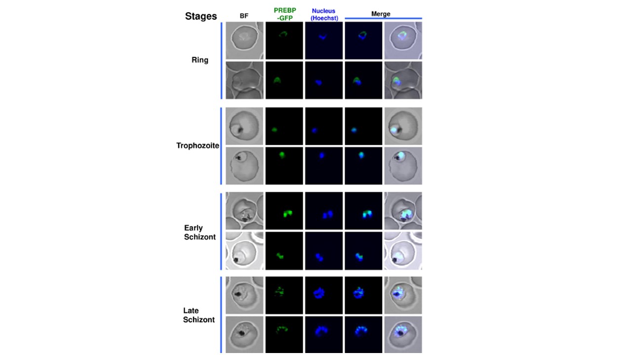
Cellular localization of PREBP-GFP fusion protein in P. falciparum cells during the intraerythrocytic stage. Living parasites that expressed PREBP-GFP fusion protein were stained with Hoechst 33342 for visualization of the nucleus. Ring, trophozoite, early schizont (with 2–3 numbers of the nucleus), and late schizont-stage parasites were observed under 407-nm emission for the detection of Hoechst (blue color), and under 488-nm emission for the detection of PREBP-GFP (green color). “BF” indicates bright-field images. “Merge” indicates merged images of Hoechst and PREBP-GFP, or those of Hoechst, PREBP-GFP, and BF. The scale bar is the same for all images. Obermann W, Azri MFD, Konopka L, Schmidt N, Magari F, Sherman J, Silva LMR, Hermosilla C, Ludewig AH, Houhou H, Haeberlein S, Luo MY, Häcker I, Schetelig MF, Grevelding CG, Schroeder FC, Lau GSK, Taubert A, Rodriguez A, Heine A, Yeo TC, Grünweller A, Taroncher-Oldenburg G. Broad anti-pathogen potential of DEAD box RNA helicase eIF4A-targeting rocaglates. Sci Rep. 2023 Jun 8;13(1):9297. doi: 10.1038/s41598-023-35765-6. PMID: 37291191