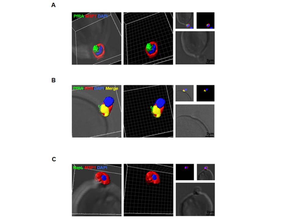
PfRA is present at the apical surface of an invading merozoite. Confocal immunofluorescence
microscopy analysis of PfRA localization in cytochalasin-D treated invading merozoites under non-permeable fixing conditions using glutaraldehyde /paraformaldehyde. (A) 3D reconstruction of z-stacks during merozoite invasion co-immunostained with MSP-1and PfRA. PfRA detected at the apical end of the merozoite surface. In the 3D images, the γ settings were altered for visual representation only. (B) PfRA (green) co-localizes with rhoptry bulb marker PfRH5 (red) that is known to be translocated to the merozoite surface to engage with its erythrocyte receptor, Basigin. (C) As a control, staining of a nuclear parasite protein, NapL, was not detected under the same non-permeable fixing conditions confirming that the fluorescent signals were obtained only from surface localized proteins. Nuclei were stained with DNA intercalating dye DAPI (Scale bar, 2 μ m.)
Anand G, Reddy KS, Pandey AK, Mian SY, Singh H, Mittal SA, Amlabu E, Bassat Q, Mayor A, Chauhan VS, Gaur D. A novel Plasmodium falciparum rhoptry associated adhesin mediates erythrocyte invasion through the sialic-acid dependent pathway. Sci Rep. 2016 6:29185.
Other associated proteins
| PFID | Formal Annotation |
|---|---|
| PF3D7_0424100 | reticulocyte binding protein homologue 5 |
| PF3D7_0930300 | merozoite surface protein 1 |
| PF3D7_1203700 | nucleosome assembly protein |
