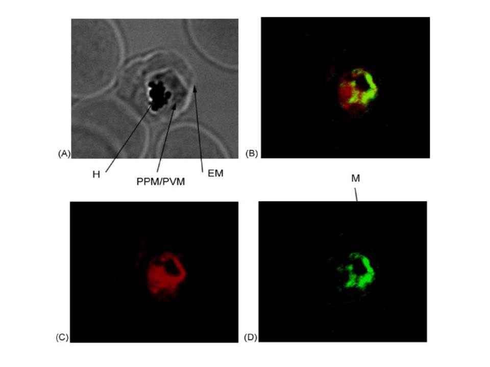
Trophozoite-infected red blood cells were probed with primary antibodies [(rabbit anti-PfIRPa 3950) and (mouse anti-Hsp60)] followed by fluorescent secondary antibodies [(goat anti-rabbit, alexa568) and (goat anti-mouse alexa488)]. The mitochondrial compartment of the intraerythrocytic trophozoite was stained in green (D), whereas PfIRPa was stained in red (C). Light microscopic image of the same trophozoite-infected red blood cell is shown in A. Colocalization of red and green fluorescence (yellow B) is indicative of a mitochondrial localization of PfIRPa, but the protein is also visualized in the parasite cytosol/food vacuole. EM—erythrocyte plasma membrane, PPM—parasite plasma membrane, PVM—parasitophorous vacuole membrane, H—hemozoin, M—mitochondrion.
Hodges M, Yikilmaz E, Patterson G, Kasvosve I, Rouault TA, Gordeuk VR, Loyevsky M. An iron regulatory-like protein expressed in Plasmodium falciparum displays aconitase activity. Mol Biochem Parasitol. 2005 143:29-38. Copyright Elsevier 2009.
Other associated proteins
| PFID | Formal Annotation |
|---|---|
| PF3D7_1342100 | aconitate hydratase |
