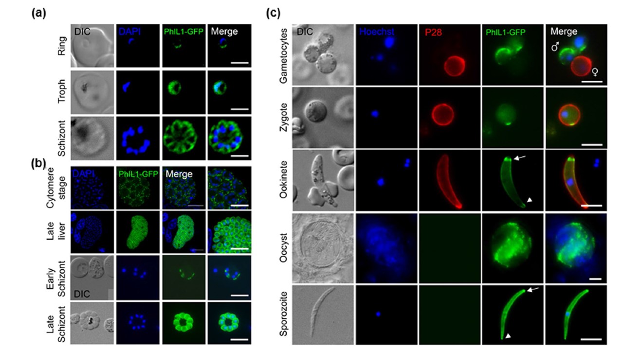
Peripheral localisation of PhIL1 during various stages of the Plasmodium life cycle as detected by live cell imaging. (a) PhIL1-GFP expression pattern in ring, trophozoite and schizont stages of P. falciparum
(b) Localization of PhIL1-GFP in early and late stages of liver and blood stage schizogony in P. berghei. (c) PhIL1-GFP expression in gametocytes, zygote, ookinete, oocyst and sporozoite stages of P. berghei. 13.1, a cy3-conjugated antibody which recognises P28 on the surface of activated females, zygotes, and ookinetes was used with the sexual stages. Arrow shows apical end and arrow head the basal end of the parasite. Scale bar = 5 μm. Schmidt S, Wichers-Misterek JS, Behrens HM, Birnbaum J, Henshall IG, Dröge J, Jonscher E, Flemming S, Castro-Peña C, Mesén-Ramírez P, Spielmann T. The Kelch13 compartment contains highly divergent vesicle trafficking proteins in malaria parasites. PLoS Pathog. 2023 19(12):e1011814. PMID: 38039338
Other associated proteins
| PFID | Formal Annotation |
|---|---|
| PF3D7_0109000 | photosensitized INA-labeled protein PHIL1 |