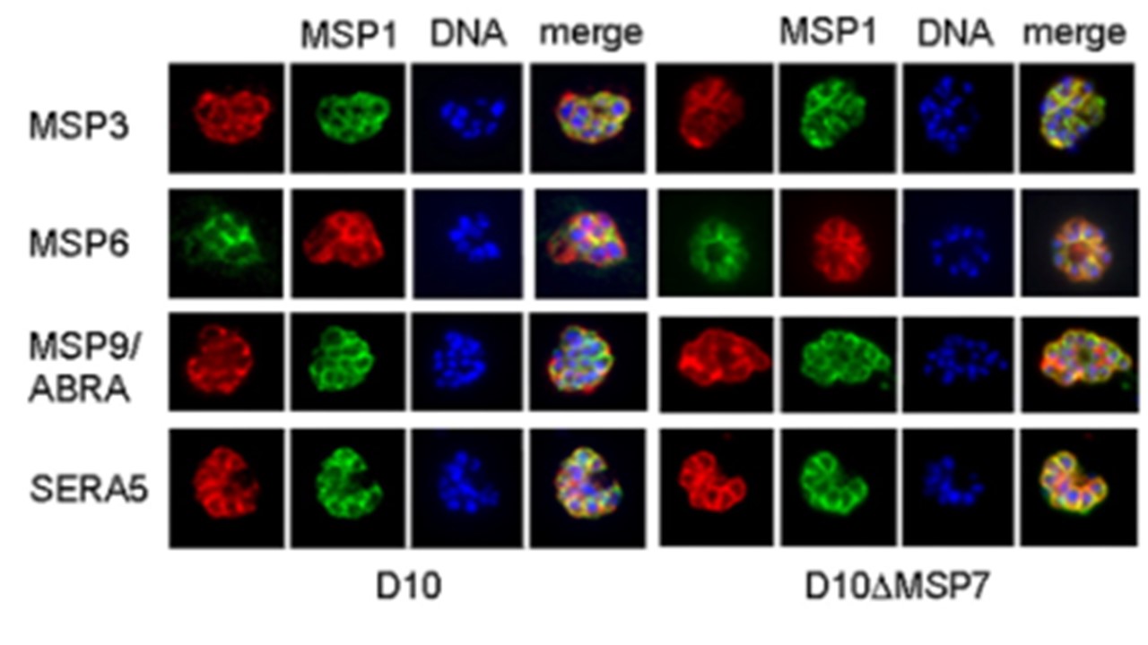
Analysis of the expression of peripheral merozoite surface and vacuolar proteins by immunoblotting and indirect immunofluorescence. (A) Immunoblot analysis of late-stage parasites (lanes 1 and 2) and purified merozoites (lanes 3 and 4) from D10 (lanes 1 and 3) or D10ΔMSP7 (lanes 2 and 4) cloned parasites with antisera to the MSP7, MSP1, MSP3, and MSP6 proteins. Antibodies to immunoglobulin binding protein (BiP) orthologue were used to ascertain that equal amounts of proteins were loaded. (B) Indirect immunofluorescence analysis of D10 and D10ΔMSP7 parasites. Fixed smears of late-stage parasites were probed with antibodies to MSP3, MSP6, MSP9, and SERA5 together with anti-MSP1 antibodies. Rabbit polyclonal antibodies are in red, while mouse antibodies are in green. Hoechst 33428 was used to visualize DNA (blue). Color-merging was carried out to analyze colocalization with MSP1 (merge). Kadekoppala M, O'Donnell RA, Grainger M, Crabb BS, Holder AA. Deletion of the Plasmodium falciparum merozoite surface protein 7 gene impairs parasite invasion of erythrocytes. Eukaryot Cell. 2008 7(12):2123-32. PMID: 18820076;
Other associated proteins
| PFID | Formal Annotation |
|---|---|
| PF3D7_0207600 | serine repeat antigen 5 |
| PF3D7_0930300 | merozoite surface protein 1 |
| PF3D7_1035400 | merozoite surface protein 3 |
| PF3D7_1228600 | merozoite surface protein 9 |
| PF3D7_1335100 | merozoite surface protein 7 |
