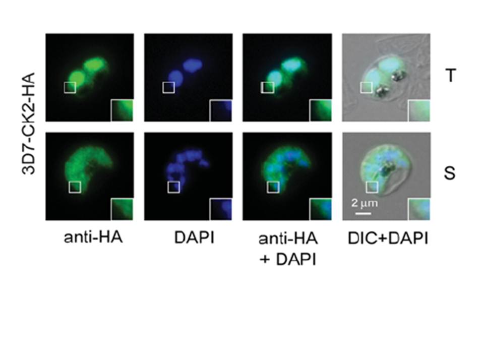
Immunofluorescence microscopy using endogenously derived Pf CK2α–HA. Localization with the anti-HA antibodies (green) confirmed expression in the nucleus (blue, DAPI) and cytosol of fixed parasites in the trophozoite (T) and schizont (S) stage. Scale bar indicates 2 μm. Enlargements of selected areas are marked with a white square. DIC, differential interference contrast microscopy. the endogenously tagged protein was localized in the parasite’s cytosol, as well as in the nucleus of trophozoite stage parasites. In the schizont stage of the parasite the dual localization of endogenously tagged CK2 was less pronounced and appeared predominantly cytosolic.
Engelberg K, Paul AS, Prinz B, Kono M, Ching W, Heincke D, Dobner T, Spielmann T, Duraisingh MT, Gilberger TW. Specific phosphoryla tion of the PfRh2b invasion
ligand of Plasmodium falciparum. Biochem J. 2013 452(3):457-66. PMID:
