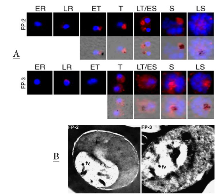
Upper panel: Localization of FP-2 and -3 by immunofluorescence across the erythrocytic life cycle: highly synchronous parasites were cultured and collected every 6 h (every 12 h for rings). Parasites were analyzed by immunofluorescence: parasites were incubated with anti-FP-2 or anti-FP-3 antisera, stained with Cy-3-conjugated secondary antibodies (red) and DAPI nuclear stain (blue), Images were merged with brightfield images to identify dark hemozoin crystals within food vacuoles (bottom panels). ER: early ring; LR: late ring; ET: early trophozoite; T: trophozoite; LT/ES: late trophozoite/early schizont; S: schizont; LS: late schizont. Falcipain-2 and -3 were localized to the food vacuole
Lower panel: FP-2 and -3 localize to the food vacuole of trophozoites: frozen thin sections of early trophozoites (for FP-2) or late trophozoites (for FP-3) were incubated with primary anti-FP2 or anti-FP3 antisera, stained with secondary antibodies-conjugated to 10nM gold beads, and evaluated by immunoelectron microscopy. Food vacuoles are labeled (fv). Arrows indicate that FP-2 and -3 is also localized in structures outside of the food vacuole.
Dahl EL, Rosenthal PJ. Biosynthesis, localization, and processing of falcipain cysteine proteases of Plasmodium falciparum. Mol Biochem Parasitol. 2005 139:205-12. Copyright Elsevier 2009.
Other associated proteins
| PFID | Formal Annotation |
|---|---|
| PF3D7_1115400 | cysteine proteinase falcipain 3 |