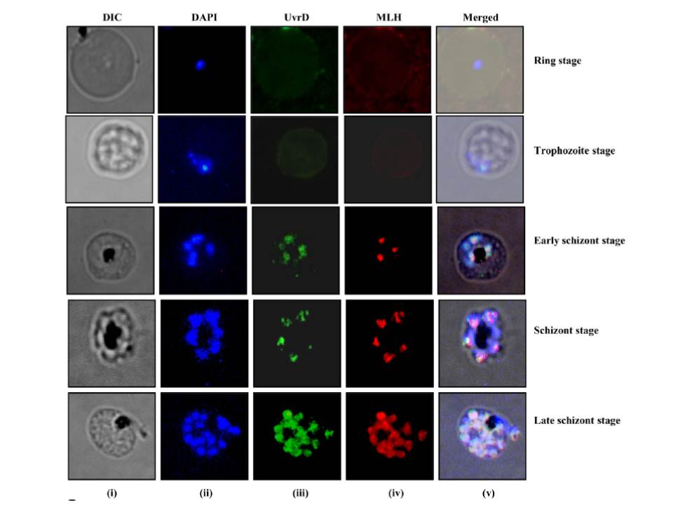
Localization of PfUvrD. Immunofluorescence staining. The cells were fixed and immunostained. Panel (i) phase (ii) image of cell stained with DAPI (iii) immunofluorescently stained cell (green, P. falciparum UvrD) (iv) immunofluorescently stained cell (red, P. falciparum MLH) and (v) super-imposed images. Both proteins co-localize in the nucleus in a cell cycle-dependent manner with peak expression in the schizont stages of the development.
Ahmad M, Ansari A, Tarique M, Satsangi AT, Tuteja R. Plasmodium falciparum UvrD Helicase Translocates in 3' to 5' Direction, Colocalizes with MLH and Modulates Its Activity through Physical Interaction. PLoS One. 2012;7(11):e49385.
Other associated proteins
| PFID | Formal Annotation |
|---|---|
| PF3D7_0514100 | ATP-dependent DNA helicase UvrD |
