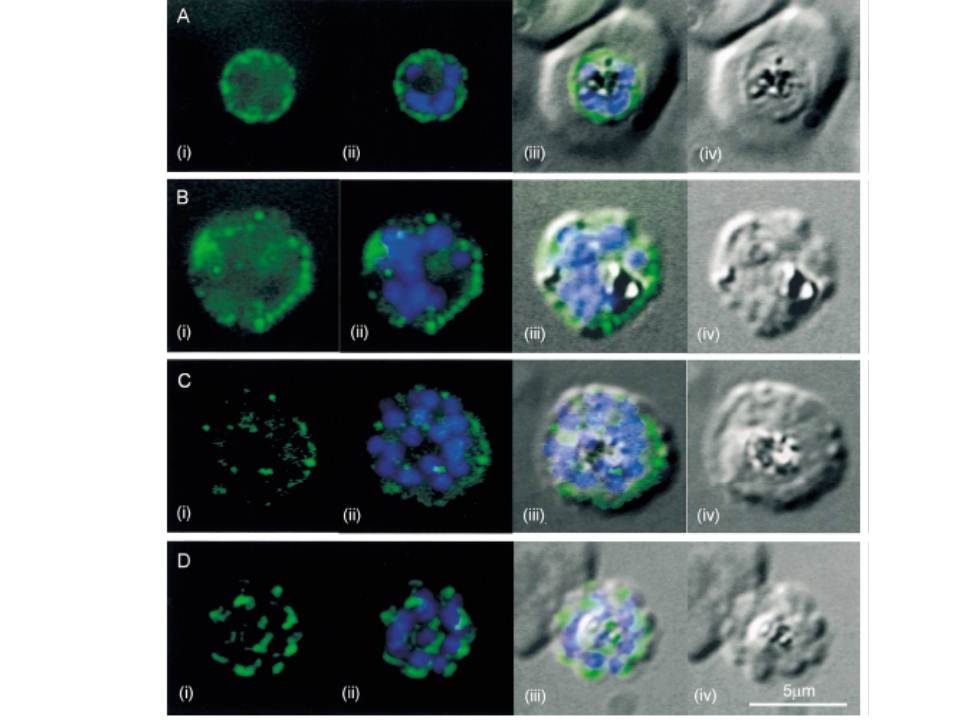
Fluorescent images of P. falciparum schizonts labelled with an antibody to cytoplasmic dynein heavy chain (green) (i), overlaid with images of the DAPI-staining of nuclei (blue) (ii), and the DIC image (iii). Plates (iv) show the DIC image alone. In plate A(i) an early schizont, with four nuclei, shows dynein labelling peripheral to the parasite nuclei (ii) and following the outline of the parasite cell body, seen in the DIC image (iv). In plate B(i) a later schizont, with 10 nuclei, shows dynein labelling, again concentrated at the periphery of the parasite. In plate C(i) a parasite with a full complement of 16 nuclei, shows less intense dynein labelling, with small points of fluorescence apparent throughout the parasite. In plate D(i) a parasite which is in the process of merozoite budding, as evident from the DIC picture (iv), and has lost its host cell membrane, shows distinct crescents of dynein labelling associated with each merozoite nucleus (ii) and the budding merozoite apices (iii).
Fowler RE, Smith AM, Whitehorn J, Williams IT, Bannister LH, Mitchell GH. Microtubule associated motor proteins of Plasmodium falciparum merozoites. Mol Biochem Parasitol. 2001 117:187-200.
Other associated proteins
| PFID | Formal Annotation |
|---|---|
| PF3D7_0718000 | dynein heavy chain, putative |
| PF3D7_0729900 | dynein heavy chain, putative |
| PF3D7_0905300 | dynein heavy chain, putative |
| PF3D7_1023100 | dynein heavy chain, putative |
| PF3D7_1202300 | dynein heavy chain, putative |
| PF3D7_1465800 | dynein beta chain, putative |
