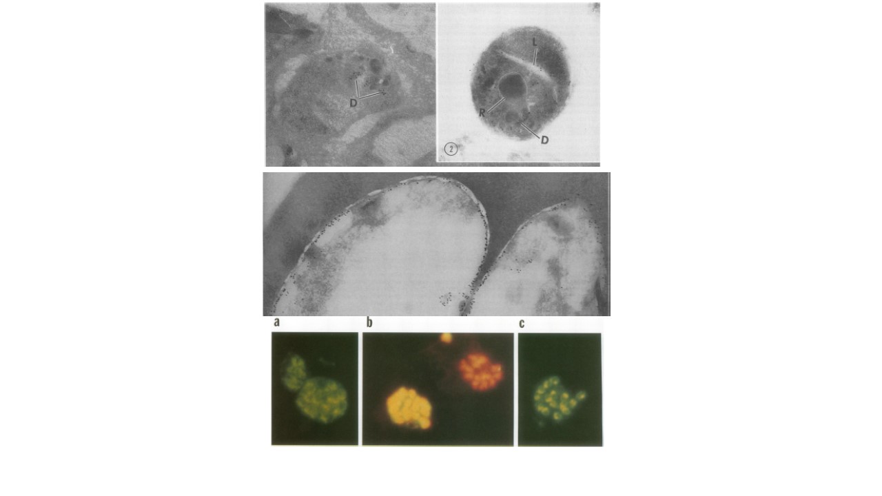
Upper panel: Section of a very late schizont, showing gold particles over the dense granules (D) of the well-formed merozoites. Note the absence of gold particles from the rhoptry (R). Magnification, x50,000. FIG. 2. Section of a free merozoite, showing gold particles at the lamellar body (L) as well as at the dense granules (D). A few gold particles are also seen at the plasma membrane. Note the absence of reaction over the flask-shaped rhoptry (R). Magnification, x65,000. Middle panel: Section through two adjacent rings, showing abundant gold particles on their plasma membranes. In contrast, very few gold particles can be seen in the immediately adjacent parasitophorous vacuole and none can be seen on the parasitophorous membrane. Magnification, x50,000. Lower panel: Immunofluorescent staining of RIMA with MAb lHl at the 8-nucleus (a) and 16-nucleus (b and c) stages. Cells in panel b were counterstained with ethidium bromide (orange) to detect nuclei. Cells staining with both fluorescein isothiocyanate and ethidium bromide appear yellow (b).
Trager W, Rozario C, Shio H, Williams J, Perkins ME. Transfer of a dense
granule protein of Plasmodium falciparum to the membrane of ring stages and
isolation of dense granules. Infect Immun. 1992 60(11):4656-61.
