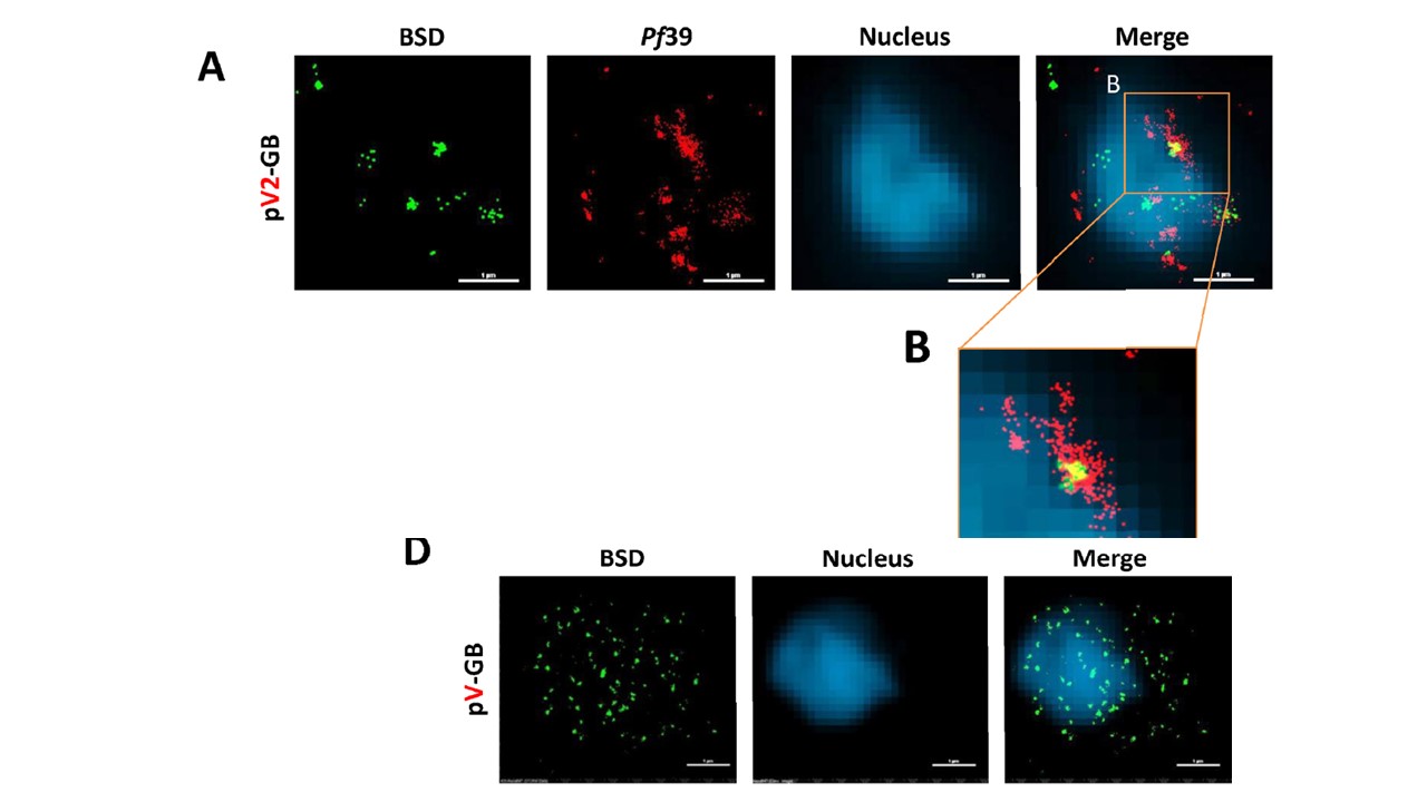
The protein expressed by the var2csa promoter is associated with the ER. (A) Super-resolution STORM imaging of parasites expressing BSDGFPfusion by a var2csa promoter (Green; pV2-GB) that were co-stained with the ER marker Pf39=ERC (red). (B) High magnification of the region indicated in orange rectangle in A. Nuclei stained with YOYO1 were imaged by conventional epi-fluorescence for cellular orientation. ( (D) Super resolution STORM imaging of parasites expressing BSD-GFP fusion by a var promoter (Green; pV-GB). Nuclei stained with YOYO1 were imaged by conventional epi-fluorescence for cellular orientation. Stochastic Optical Reconstruction Microscopy (STORM) was applied , which is a technique sensitive enough to detect photons from single molecules, and were able to detect the deaminase expressed by var2csa in small complexes localized mainly to the ER (A–B), while the protein expressed by a typical var promoter was distributed throughout the parasite cytoplasm.
Fastman Y, Assaraf S, Rose M, Milrot E, Basore K, Arasu BS, Desai SA, Elbaum M, Dzikowski R. An upstream open reading frame (uORF) signals for cellular localization of the virulence factor implicated in pregnancy associated malaria. Nucleic Acids Res. 2018 [Epub ahead of print]
Other associated proteins
| PFID | Formal Annotation |
|---|---|
| PF3D7_1108600 | endoplasmic reticulum-resident calcium binding protein |
