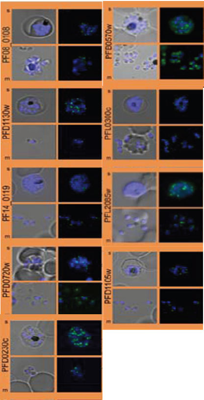
Subcellular distribution of proteins predicted to be involved in invasion. All proteins were localized in schizonts (s) and free merozoites (m) using GFP-fusion proteins and grouped according to their predominant GFP localization. Apical. Nuclei were stained with DAPI (blue).
Hu G, Cabrera A, Kono M, Mok S, Chaal BK, Haase S, Engelberg K, Cheemadan S, Spielmann T, Preiser PR, Gilberger TW, Bozdech Z. Transcriptional profiling of growth perturbations of the human malaria parasite Plasmodium falciparum. Nat Biotechnol. 2010 28(1):91-8.
PubMed Article: Transcriptional profiling of growth perturbations of the human malaria parasite Plasmodium falciparum
Other associated proteins
| PFID | Formal Annotation |
|---|---|
| PF3D7_0212600 | secreted protein with altered thrombospondin repeat domain |
| PF3D7_0404700 | dipeptidyl aminopeptidase 3 |
| PF3D7_0414900 | armadillo-domain containing rhoptry protein |
| PF3D7_0423400 | apical asparagine-rich protein AARP |
| PF3D7_0423800 | RH5-Ripr membrane anchoring protein cysteine-rich protective antigen |
| PF3D7_0808700 | erythrocyte membrane protein 1, PfEMP1 |
| PF3D7_1412000 | p1/s1 nuclease, putative |
