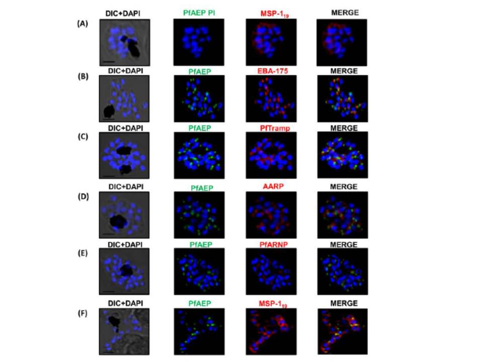
PfAEP localization in schizont stages analyzed by confocal microscopy. Subcellular localization of PfAEP was studied by co-staining with antibodies against micronemal protein EBA-175 (B), rhoptry bulb protein PTRAMP (C), and rhoptry neck,
apical asparagine rich protein (AARP) (D), apical rhoptry neck protein (ARNP) (E), and merozoite surface protein (MSP) (F).As a control, pre-immune serum of PfAEP was checked and no staining was observed in schizonts (A). Mature schizonts were costained with anti-PfAEP (green) and anti-EBA-175, TRAMP, AARP, ARNP, MSP-119 antibodies (red). The nuclei of schizonts were stained with DAPI (blue) and visualized by Nikon N-SIM confocal microscope. All apical marker proteins and PfAEP showed punctate staining in schizonts distinct from DAPI. Co-staining of PfAEP with surface marker MSP-119 showed PfARNP localized at the apical tip with surface of merozoites stained by MSP-119. PfAEP staining did not merge with either markers of microneme or rhoptry indicating that PfAEP is neither a resident of micronemes or rhoptry of
merozoites. Scale bar 2 mm.
Hans N, Relan U, Dubey N, Gaur D, Chauhan VS. Identification and localization of a Novel Invasin of Plasmodium falciparum. Mol Biochem Parasitol. 2015 Sep 29. [Epub ahead of print]
Other associated proteins
| PFID | Formal Annotation |
|---|---|
| PF3D7_0423400 | apical asparagine-rich protein AARP |
| PF3D7_0511600 | apical rhoptry neck protein |
| PF3D7_0731500 | erythrocyte binding antigen-175 |
| PF3D7_0930300 | merozoite surface protein 1 |
| PF3D7_1137200 | basal complex transmembrane protein 1 |
