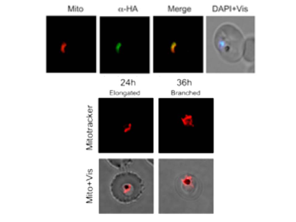
Upper panel: Immunofluorescence assay showing transgenic parasites expressing PfClpQ(mut)-HA-ddFKBP (ClpQ(mut)-DD parasite line) immuno-stained with anti-HA antibodies and co-stained for mitochondria with MitoTracker. The parasite nuclei were stained with DAPI and slides were visualized by fluorescence microscope. the immunofluorescence staining overlapped with the MitoTracker staining suggesting that the fusion protein is localized in the mitochondria which is the site of action of native protein
Lower panel: Fluorescent microscopic images of MitoTracker stained ClpQ(mut)-DD transgenic parasites at different time points (lower panel). The mitochondria in control ClpQ(mut)-DD set showed normal growth and development; in majority of these parasites at trophozoite stage the mitochondria appeared as elongated structures, in late trophozoite/early schizont stage) most of these mitochondria showed branched structure.
Jain S, Rathore S, Asad M, Hossain ME, Sinha D, Datta G, Mohmmed A. The prokaryotic ClpQ protease plays a key role in growth and development of mitochondria in Plasmodium falciparum. Cell Microbiol. 2013 15(10):1660-73
