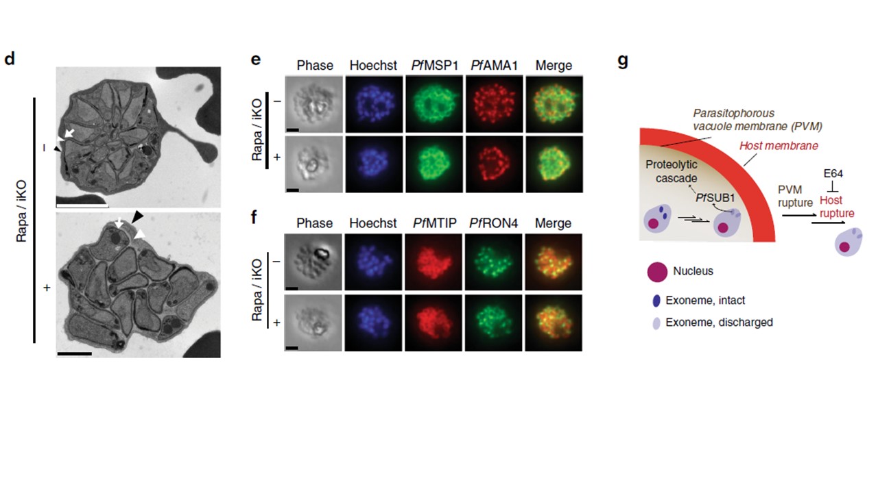
PfPP1 function at an early step of parasite egress from erythrocytes. d Electron microscopy of terminally developed pfpp1-iKO parasites treated +/−Rapa at 30-hpi. In both images, the different membranes are indicated as follows: erythrocyte (black arrowhead), PV (white arrowhead), and parasite (white arrow). Representative of 2 experiments. Scale bars: 2 μm (top), 1 μm (bottom). e, f Immunofluorescence analysis of the microneme antigen PfAMA1 e or the rhoptry-neck antigen PfRON4 f in terminally developed parasites +/−iKO of pfpp1 at 30 hpi. The images also show the parasite plasma membrane marker PfMSP1 e and the inner membrane complex marker PfMTIP f. Scale bar: 2 μm. For both panels, representative of 2 experiments. g In a mature parasite, regulated secretion of PfSUB1 from exonemes stimulates a proteolytic cascade leading to sequential rupture of the PVM and the erythrocyte host membranes.
Paul AS, Miliu A, Paulo JA, Goldberg JM, Bonilla AM, Berry L, Seveno M, Braun-Breton C, Kosber AL, Elsworth B, Arriola JSN, Lebrun M, Gygi SP, Lamarque MH, Duraisingh MT. Co-option of Plasmodium falciparum PP1 for egress from host erythrocytes. Nat Commun. 2020 11(1):3532.
Other associated proteins
| PFID | Formal Annotation |
|---|---|
| PF3D7_0930300 | merozoite surface protein 1 |
| PF3D7_1116000 | rhoptry neck protein 4 |
| PF3D7_1133400 | apical membrane antigen 1 |
| PF3D7_1414400 | serine/threonine protein phosphatase PP1 |
