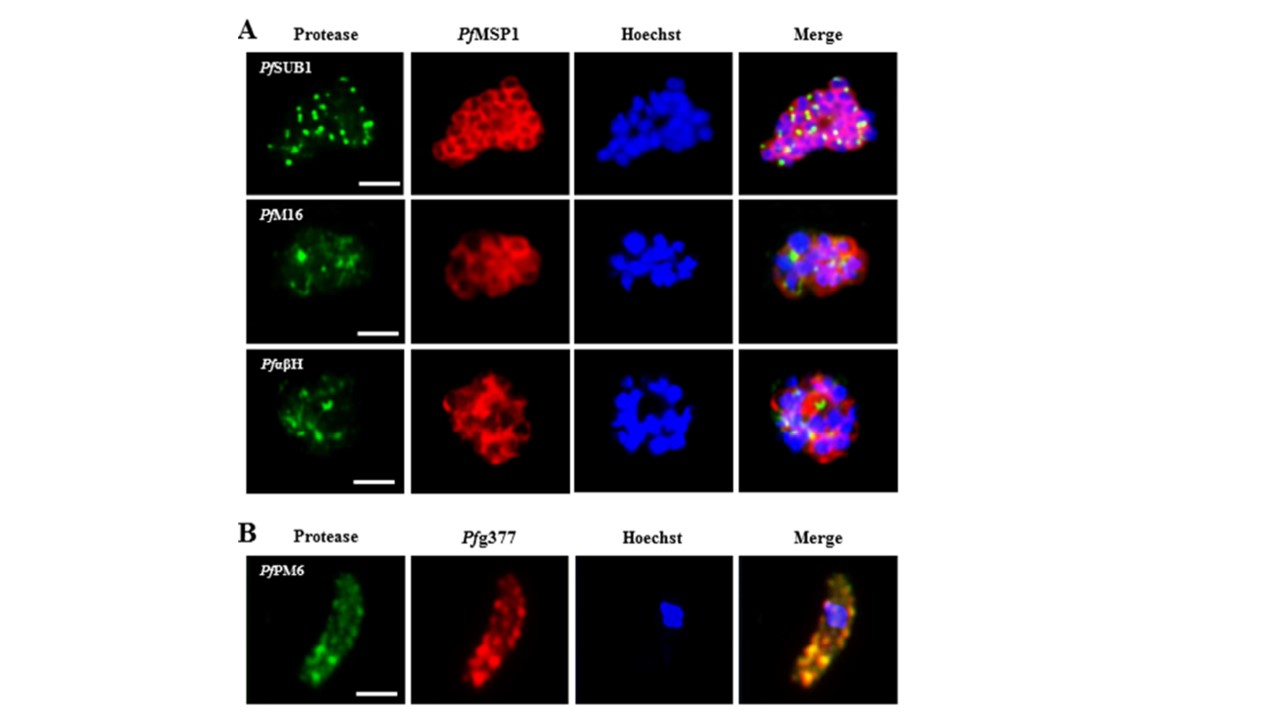
In-depth analysis of protease localization. A) IFAs using polyclonal mouse antisera directed against PfSUB1, PfαβH and PfM16 were used to investigate the granular localization of these proteases in merozoite-containing mature schizonts (in green). The merozoites were visualized via immunolabelling with polyclonal rabbit antisera directed against PfMSP1 (in red). PfM16 localized to granular structures in schizonts, which did not coincide with the merozoites B) Immunolabeling of osmiophilic bodies using polyclonal rabbit antisera directed against Pfg377 (in red) was employed to demonstrate an accumulation of PfPM6, labeled with the respective mouse antisera (in green), in these organelles. Hoechst 33342 staining was used to highlight the parasite nuclei (in blue). Bar, 2 μm. Results are representative of two independent experiments. The localization of PfPM6 in female gametocytes was don by co-localization experiments were performed, using rabbit antibodies directed against the osmiophilic body protein Pfg377. The double-labelling assays demonstrated that PfPM6 is present in these organelles.
Weißbach T, Golzmann A, Bennink S, Pradel G, Julius Ngwa C. Transcript and protein expression analysis of proteases in the blood stages of Plasmodium falciparum. Exp Parasitol. 2017 Mar 25. [Epub ahead of print]
Other associated proteins
| PFID | Formal Annotation |
|---|---|
| PF3D7_0311700 | plasmepsin VI |
| PF3D7_0507500 | subtilisin-like protease 1 |
| PF3D7_0930300 | merozoite surface protein 1 |
| PF3D7_1121800 | peptidase m16, putative |
| PF3D7_1410100 | alpha/beta hydrolase, putative |
