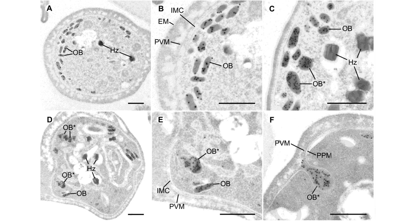
(A-B) An overview image (A) and its magnified image (B) of a non-activated female gametocyte of P. falciparum. Gold particles indicating the localization of PfG377 are observed in osmiophilic bodies (OB) of a non-activated female gametocyte. Hz, hemozoin granule; IMC, inner membrane complex; PVM, parasitophorous vacuole membrane; EM, erythrocyte membrane. (C-F) Activated female gametocytes after 30 min incubation in activation medium. Several osmiophilic bodies fuse to form larger vesicles (OB*). An overview image of an activated female gametocyte (D) and enlargement showing the lower left part (E). Vesicles formed by fusion of osmiophilic bodies (OB*) are found near the IMC of an activated female gametocyte. (F) Membrane of a large vesicle (OB*) is fused with parasite plasma membrane (PPM), and the contents of the vesicle are ready to be secreted into the PV space or the PVM.
Ishino T, Tachibana M, Baba M, Iriko H, Tsuboi T, Torii M. Observation of morphological changes of female osmiophilic bodies prior to Plasmodium gametocyte egress from erythrocytes. Mol Biochem Parasitol. 2020 Jan 22:111261.
