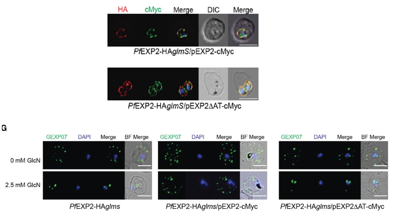
Complementation of PfEXP2-HAglmS with episomally expressed truncated EXP2ΔAT-cMyc. (E) IFA of infected erythrocytes with DAPI staining (blue) marking the parasite’s nucleus (left), and the degree of co-localization between endogenous EXP2 (HA) and episomally expressed cMyc-tagged EXP2 calculated by measuring Pearson’s coefficients of merged Z-stack images of 20 cells (right). Shown is the mean ± SD, with significance determined by an unpaired t-test (****p < 0.001. (G) IFA of infected erythrocytes using antibodies to the Maurer’s cleft protein GEXP07 and quantification of GEXP07 localization (n = 30 cells from a single experiment), defined as either fully exported, not exported, or showing an intermediate export phenotype. Scale bar, 5 μm. Pitman EL, Counihan NA, Modak JK, Chowdury M, Gilson PR, Webb CT, de Koning-Ward TF. Dissecting EXP2 sequence requirements for protein export in malaria parasites. Front Cell Infect Microbiol. 2024 13:1332146. PMID: 38282616
Other associated proteins
| PFID | Formal Annotation |
|---|---|
| PF3D7_1471100 | exported protein 2 |
