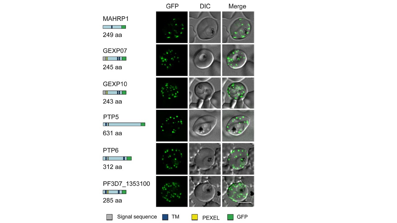
Live-cell fluorescence analysis of GFP-tagged exported proteins. Predicted native protein lengths (amino acids [aa]) and schematic representations of six proteins that were selected for GFP tagging are shown on the left. Gray = signal sequence, blue = transmembrane domain (TM), yellow = PEXEL motif, green = GFP tag. Live-cell fluorescence and DIC microscopy of transfectants expressing GFP-tagged putative Maurer’s cleft proteins revealed fluorescent puncta in the RBC cytoplasm. Scale bar=5 mm. we visualized each of the GFP-tagged proteins as puncta in the host RBC cytoplasm, consistent with a Maurer’s cleft association.
McHugh E, Carmo OMS, Blanch A, Looker O, Liu B, Tiash S, Andrew D, Batinovic S, Low AJY, Cho HJ, McMillan P, Tilley L, Dixon MWA. Role of Plasmodium falciparum Protein GEXP07 in Maurer's Cleft Morphology, Knob Architecture, and P. falciparum EMP1 Trafficking. mBio. 2020 11(2). pii: e03320-19.
Other associated proteins
| PFID | Formal Annotation |
|---|---|
| PF3D7_0113900 | Plasmodium exported protein (hyp8) |
| PF3D7_1002100 | EMP1-trafficking protein |
| PF3D7_1302000 | EMP1-trafficking protein |
| PF3D7_1353100 | Plasmodium exported protein, unknown function |
| PF3D7_1370300 | membrane associated histidine-rich protein |
