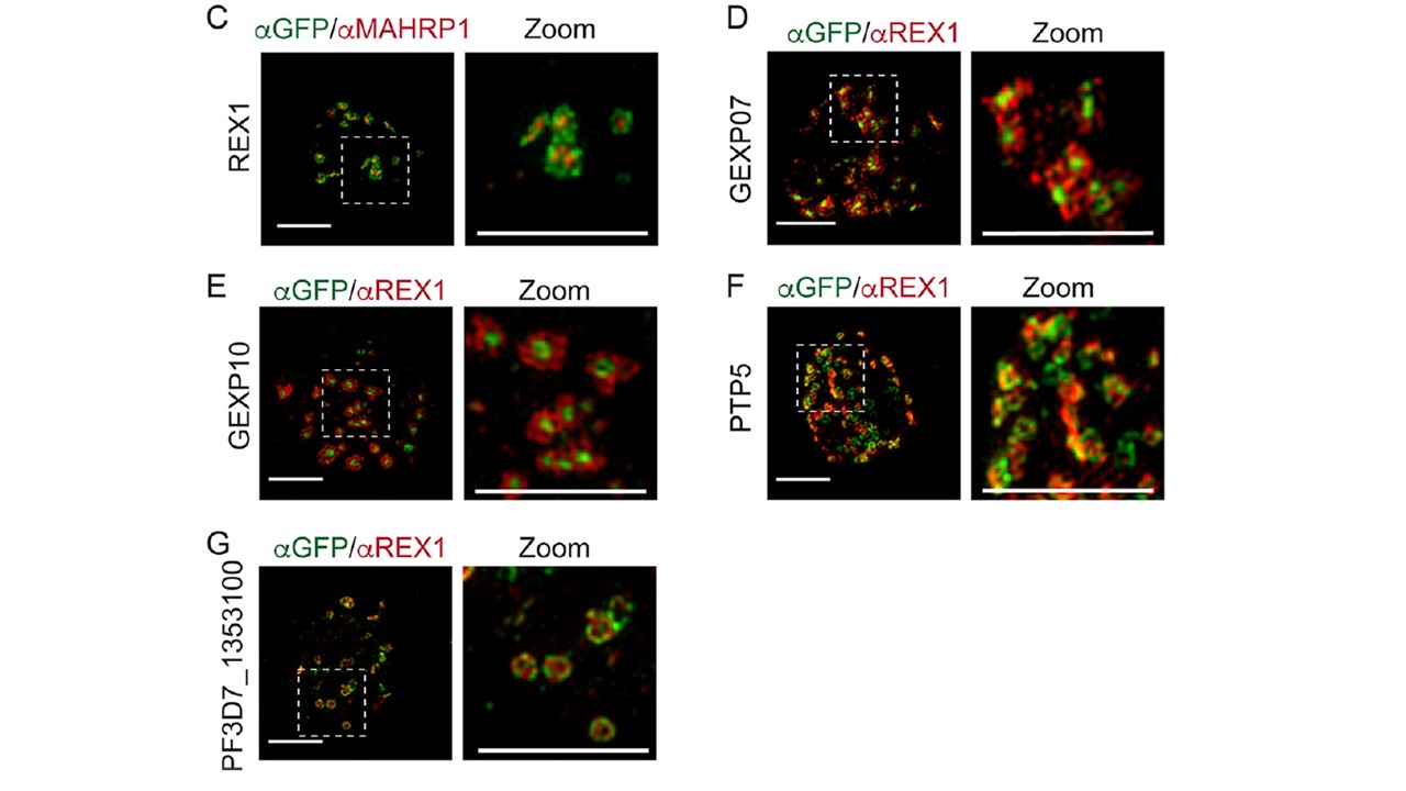
Maurer’s cleft proteins interact to form distinct protein clusters. (C) 3D-SIM analysis of REX1-GFP-infected RBCs fixed and labeled with anti-GFP (green) and anti-MAHRP1 (red). (D to G) 3D-SIM analysis of transfectant-infected RBCs expressing GFP-tagged GEXP07, GEXP10, PTP5, and Pf3D7_1353100 that were fixed and labeled with anti-GFP (green) and anti-REX (red) antibodies. Maximum projections of Z-stacks are displayed. Scale bars=3 mm, zoom scale Bar=3 mm. 3D-SIM revealed that REX1 was located around the perimeter of the clefts, while MAHRP1 exhibited a more central location (C). GEXP07-GFP and GEXP10-GFP are concentrated in the central region of the Maurer’s cleft surrounded by REX1 (D). 3D-SIM imaging of PTP5-GFP and PF3D7_1353100-GFP cell lines revealed a dotted pattern at the periphery of the Maurer’s cleft cisternae, partially overlapping or alternating with the REX1 signal (F and G). Similarly, PfEMP1 (ATS labeling) partly overlapped REX1 and PfEMP1 at the cleft periphery,
McHugh E, Carmo OMS, Blanch A, Looker O, Liu B, Tiash S, Andrew D, Batinovic S, Low AJY, Cho HJ, McMillan P, Tilley L, Dixon MWA. Role of Plasmodium falciparum Protein GEXP07 in Maurer's Cleft Morphology, Knob Architecture, and P. falciparum EMP1 Trafficking. mBio. 2020 11(2). pii: e03320-19.
Other associated proteins
| PFID | Formal Annotation |
|---|---|
| PF3D7_0113900 | Plasmodium exported protein (hyp8) |
| PF3D7_0935900 | ring-exported protein 1 |
| PF3D7_1353100 | Plasmodium exported protein, unknown function |
| PF3D7_1370300 | membrane associated histidine-rich protein |
