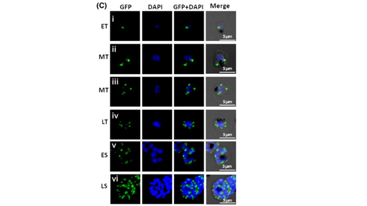
Localization of Pfμ1 in different intracellular stages of
(A) Schematic diagram of the wild type Pfμ1 (PF3D7_1311400) P. falciparum. showing location of β-binding domain and cargo-binding domain. The complete gene was cloned in frame with GFP in the pARL1a + vector under the control of chloroquine resistant transporter gene promoter (crt 5′ UTR) and dhfr terminator (3′ UTR). (B i) Immunoblot analysis of whole cell lysates of trophozoite-stage 3D7 and transgenic parasites expressing Pfμ1–GFP by α-GFP antibody shows a band at ~ 76 kDa. (B ii) Immunoblot analysis of whole cell lysates of trophozoite-stage 3D7 and transgenic parasites expressing Pfμ1–GFP by α-Pfμ1 antibody. A band at ~ 50 kDa, representing the native protein is recognized by the Pfμ1 antibody in lanes 1 and 2, while another band at ~ 76 kDa (lane 2), representing the Pfμ1–GFP fusion, was recognized in the transgenic line only. (B iii) Loading control lane as probed by anti-Pf BiP. (C) Live cell imaging of transgenic parasites expressing the Pfμ1–GFP fusion protein from early trophozoite to late schizont stages. Parasite nuclei were stained with DAPI; scale bars denote 5 μM. ET, Early Trophozoite; MT, Mid Trophozoite; LT, Late Trophozoite; ES, Early Schizont; LS, Late Schizont. Kaderi Kibria KM, Rawat K, Klinger CM, Datta G, Panchal M, Singh S, Iyer GR, Kaur I, Sharma V, Dacks JB, Mohmmed A, Malhotra P. A role for adaptor protein complex 1 in protein targeting to rhoptry organelles in Plasmodium falciparum. Biochim Biophys Acta. 2015 1853(3):699-710. PMID: 25573429.
