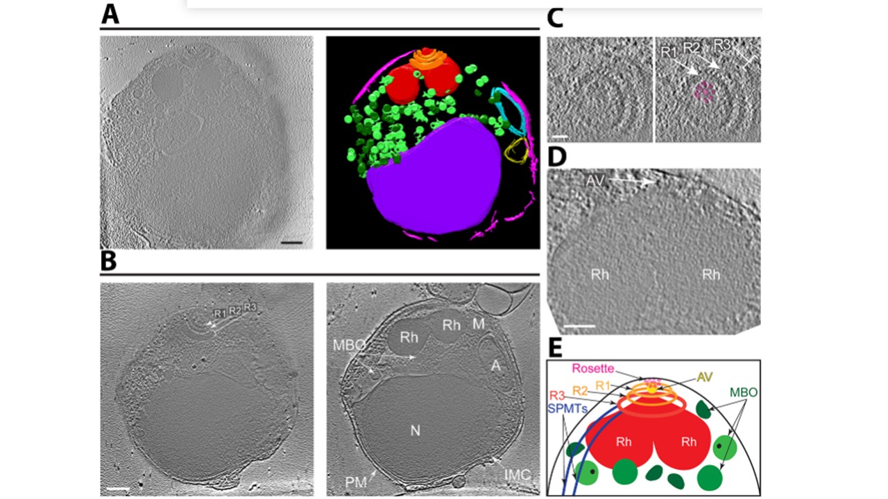
Cryo-ET with Volta Phase Plate reveals the organization of subcellular organelles in Plasmodium falciparum merozoites. (A) Tomographic slice of a representative merozoite (left) and its 3D annotation showing the apical rings (orange), rhoptries (red), membrane-bound organelles (light/dark green), inner membrane complex (pink), apicoplast (cyan), mitochondrion (yellow, presumptively designated based on double-membrane structure and position adjacent to the four-membrane apicoplast), and nucleus (purple). Scale bar, 200 nm. Movie with the complete tomogram and annotation is in Video S1. (B) Two tomographic slices of a merozoite (different from that shown in [A]) that better show the three apical rings (Ring 1 [R1], Ring 2 [R2], Ring 3 [R3]), rhoptries (Rh), membrane-bound organelles (MBO), inner membrane complex (IMC), apicoplast (A), mitochondrion (M), nucleus (N), and plasma membrane (PM). Scale bar, 200 nm. (C) Zoomed-in view of a tomographic slice of a merozoite (different from [A] and [B]) showing the apical rings (Rings 1 and 2, arrows; Ring 3, bracket) and the rosette (highlighted in magenta). Scale bar, 50 nm. (D) Zoomed-in view of a tomographic slice of a merozoite (different from [A], [B], and [C]) showing the apical vesicle (AV; arrow) at the tip of two rhoptries (R). Scale bar, 100 nm. (E) Cartoon of the Plasmodium merozoite apical complex showing the arrangement of key subcellular structures. Abbreviations as in (B) except subpellicular microtubules (SPMTs) and apical vesicle (AV) are also indicated. Sun SY, Segev-Zarko L-a, Pintilie GD, Kim CY, Staggers SR, Schmid MF, Egan ES, Chiu W, Boothroyd JC. Cryogenic electron tomography reveals novel structures in the apical complex of Plasmodium falciparum. mBio. 2024 e0286423. PMID: 38456679.