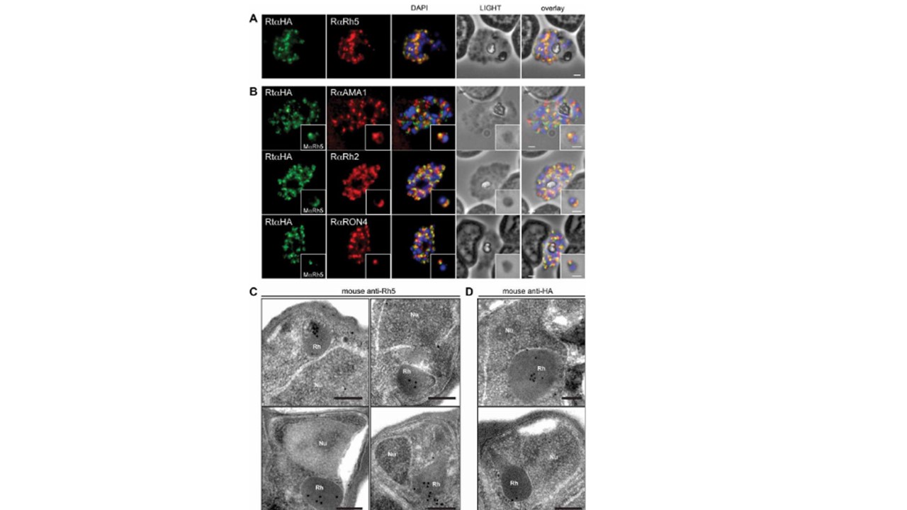
PfRh5 localises to the rhoptry body. (A) Immunofluorescence and phase contrast micrographs of late segmented schizonts with anti-HA antibodies to detect the
chimeric PfRh5 along with co-localisation using antibodies to PfRh5. The panels from left to right are rat anti-HA, rabbit anti-Rh5, overlay of both with DAPI nuclear stain, phase contrast image and overlay of all images. Scale bars = 1 lM. (B) Immunofluorescence and phase contrast images of late schizonts or free merozoites (insets) to co-localise PfRh5 with AMA1 (top panel), PfRh2a/b (middle panel) and RON4 (bottom panel). Each panel from left top right corresponds to anti-HA antibodies (to detect PhRh5), rabbit anti-AMA1 or anti-Rh2a/b or anti-RON4, overlay of each with DAPI nuclear stain, phase contrast and overlay of all images. Insets show individual merozoites. Scale bars = 1 mm. (C) Immuno-electron microscopy of late schizonts localising PfRh5 (using both anti-PfRh5 and anti-HA in the tagged line) to the merozoite rhoptries. Scale bars = 0.2 mm. Triglia T, Tham WH, Hodder A, Cowman AF. Reticulocyte binding protein homologues are key adhesins during erythrocyte invasion by Plasmodium falciparum. Cell Microbiol. 2009
Other associated proteins
| PFID | Formal Annotation |
|---|---|
| PF3D7_0424100 | reticulocyte binding protein homologue 5 |
| PF3D7_1116000 | rhoptry neck protein 4 |
| PF3D7_1133400 | apical membrane antigen 1 |
| PF3D7_1335400 | reticulocyte binding protein 2 homologue a |