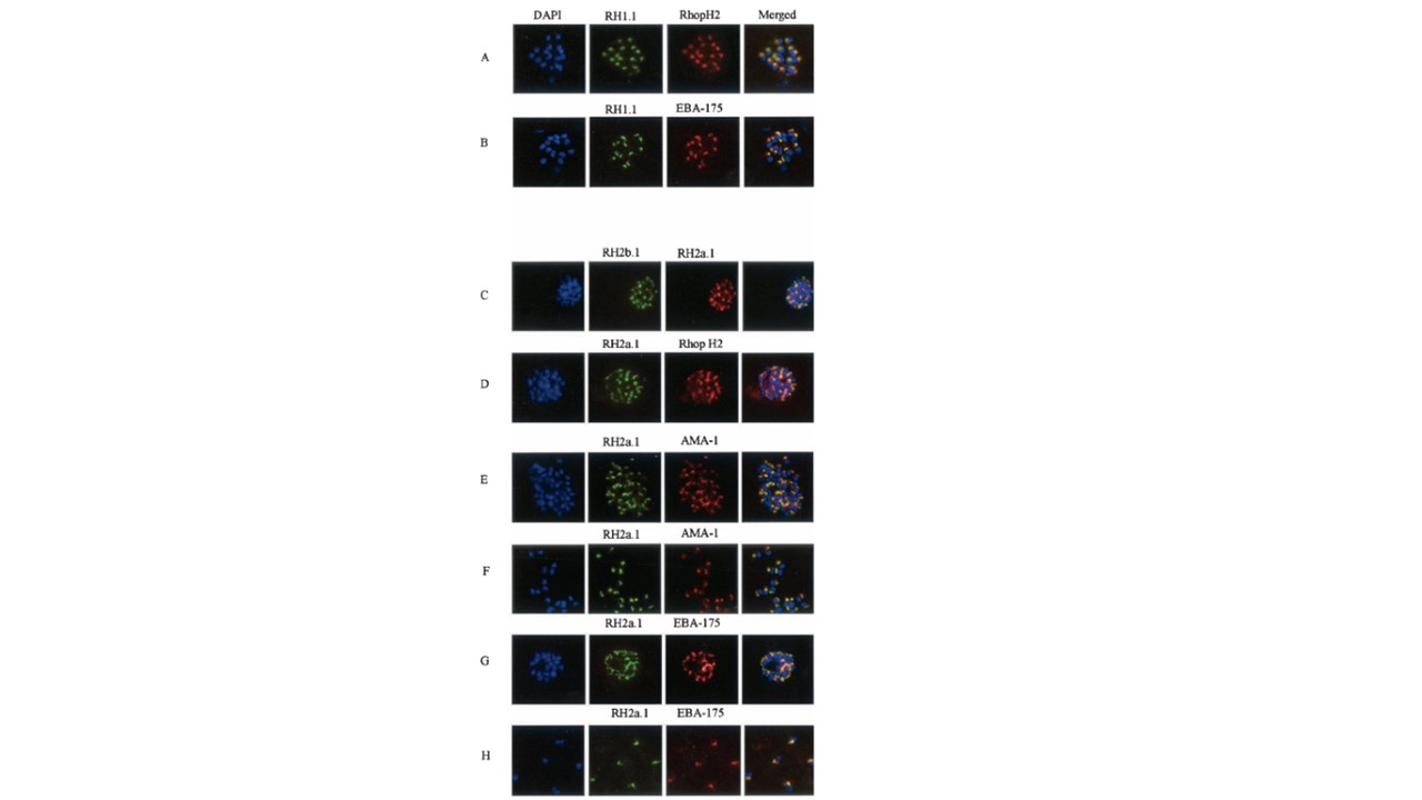
Double-staining IFA of FCB1 and 3D7 parasites. (A and B) FCB1 parasites. (C to H) 3D7 parasites. All panels show late schizonts, except for panels F and H, which show released merozoites. The following primary antibodies were used: mouse polyclonal antisera RH1.1, RH2a.1, and RH2b.1; rabbit polyclonal antiserum RH2a.1 and antiserum to RhopH2 (a rhoptry protein); rat polyclonal antiserum to EBA-175 (a microneme protein); and rat MAb 4G2dc1 to AMA-1 (thought to be located in the rhoptries). Parasite nuclei are stained with DAPI (blue). For each panel, the FITC-conjugated secondary antibodies (green) are to mouse polyclonal sera, and the TRITC-conjugated secondary antibodies (red) are to rabbit polyclonal sera, with the following exceptions. (E and F) Rabbit antiserum RH2a.1 was labeled with FITC-conjugated anti-rabbit immunoglobulin G (IgG) (Sigma), and rat MAb 4G2dc1 was labeled with Texas red-conjugated anti-rat IgG (Sigma). (H) Rabbit antiserum RH2a.1 was labeled as described above, and rat antiserum to EBA-175 was
labeled with Texas red-conjugated anti-rat IgG. In the merged images, areas of overlap between the red and the green signals are shown in yellow. Scale bar, 10 mm. Taylor HM, Grainger M, Holder AA. Variation in the expression of a Plasmodium falciparum protein family implicated in erythrocyte invasion. Infect Immun. 2002. PMID: 12228308
Other associated proteins
| PFID | Formal Annotation |
|---|---|
| PF3D7_0402300 | reticulocyte binding protein homologue 1 normocyte binding protein 1 |
| PF3D7_0731500 | erythrocyte binding antigen-175 |
| PF3D7_0929400 | high molecular weight rhoptry protein 2 |
| PF3D7_1133400 | apical membrane antigen 1 |
| PF3D7_1335400 | reticulocyte binding protein 2 homologue a |