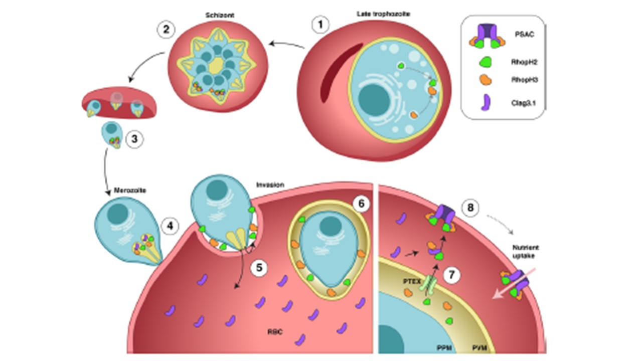
Model for RhopH proteins trafficking onto the RBC surface. a RhopH2 and RhopH3 colocalize in late schizonts (1 and 2). Upon egress (3) free
merozoites attach to new red blood cells (4). During invasion, rhoptry content is discharged and RhopH2 and RhopH3 are released into the nascent parasitophorous vacuole while Clag3 is predominantly injected into the host cell cytoplasm (5). Upon successful invasion, RhopH2 and RhopH3 are confined within the parasitophorous vacuole but show very little interaction (6). In trophozoites, RhopH2 and RhopH3 are exported via the PTEX translocon (7). In the host cell cytoplasm, both RhopH2 and RhopH3 associate with Clag3 and the RhopH complex consisting of RhopH2, RhopH3 and Clag3 can spontaneously insert into the erythrocyte membrane, where it forms the nutrient channel (8). Pasternak M, Verhoef JMJ, Wong W, Triglia T, Mlodzianoski MJ, Geoghegan N, Evelyn C, Wardak AZ, Rogers K, Cowman AF. RhopH2 and RhopH3 export enables assembly of the RhopH complex on P. falciparum-infected erythrocyte membranes. Commun Biol. 2022 5(1):333. PMID: 35393572
Other associated proteins
| PFID | Formal Annotation |
|---|---|
| PF3D7_0302200 | cytoadherence linked asexual protein 3.2 |
| PF3D7_0905400 | high molecular weight rhoptry protein 3 |
| PF3D7_0929400 | high molecular weight rhoptry protein 2 |
