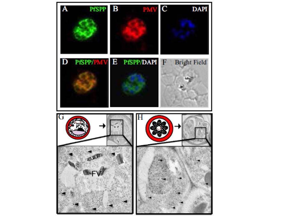
Co-localization of PfSPP (signal peptide peptidase) and Plasmepsin V in the endoplasmic reticulum. Indirect immunofluorescence microscopy using specific antibodies demonstrates PfSPP (green) co-localizes with a known parasite ER marker Plasmepsin V (red) in the Plasmodium falciparum (3D7) trophozoite. (A) Staining of PfSPP. (B) Staining of known ER marker, Plasmepsin V. (C) DAPI stain. (D) PfSPP and PMV overlay. (E) Overlay of PfSPP and DAPI stain. (F) Bright field microscopy. (G and H) Immunogold electron microscopy of PfSPP in the parasite infected erythrocytes. PfSPP is indicated by black dots and several spots are identified by black arrowheads. (G) Schematic illustration of a mature trophozoite stage parasite in the infected. PfSPP staining in the trophozoite shows a diffuse staining pattern throughout the perinuclear endomembrane system/ER. (H) Illustration of a late schizont-stage parasite in an infected erythrocyte. PfSPP staining in the late-schizont stage of parasite development shows staining throughout the perinuclear endomembrane system/ER.
Baldwin M, Russo C, Li X, Chishti AH. Plasmodium falciparum signal peptide peptidase cleaves malaria heat shock protein 101 (HSP101). Implications for gametocytogenesis. Biochem Biophys Res Commun. 2014 Jul 10 [Epub ahead of print]
Other associated proteins
| PFID | Formal Annotation |
|---|---|
| PF3D7_1323500 | PEXEL protease plasmepsin V |
