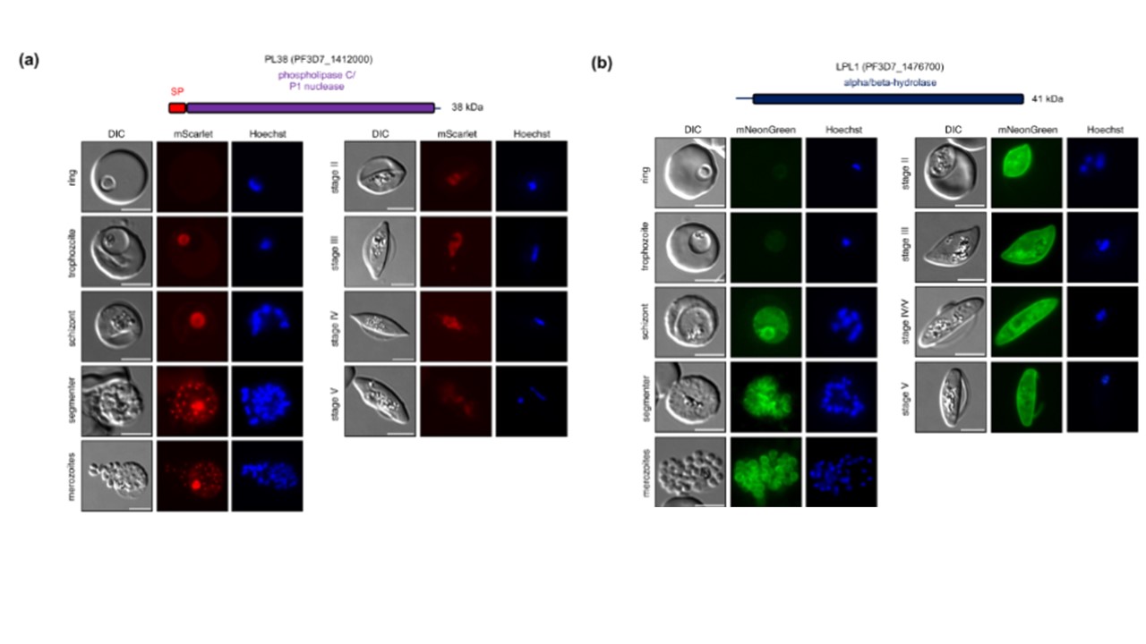
Localization analysis of PL38 and LPL1. Parasites expressing endogenously tagged PL38-mScarlet (a) and LPL1-mNeonGreen (b) were analyzed during asexual and sexual blood stage development by live-cell microscopy. Nuclei were stained with Hoechst. Scale bars = 5 μm. DIC, differential interference contrast. Schematic representations of the functional domains of the two proteins are shown on top of the images. SP, signal peptide.
Pietsch E, Niedermüller K, Andrews M, Meyer BS, Lenz TL, Wilson DW, Gilberger TW, Burda PC. Disruption of a Plasmodium falciparum patatin-like phospholipase delays male gametocyte exflagellation. Mol Microbiol. 2023 PMID: 38131156.
PubMed Article: Disruption of a Plasmodium falciparum patatin-like phospholipase delays male gametocyte exflagellation
Other associated proteins
| PFID | Formal Annotation |
|---|---|
| PF3D7_1412000 | p1/s1 nuclease, putative |
