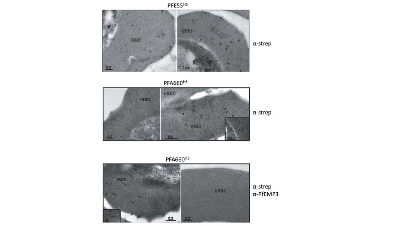
Transmission electron microscopy of immunogold labelling of PFE55HS (upper panel), PFA660HS (middle and lower panels) using anti-strep antibodies (large gold particles). In the lower panel co-labelling was carried out with anti-PfEMP3 antisera (small gold particles). As a control for antibody specificity, lower panel (right) shows a non-infected erythrocyte treated under the same conditions. Gold particles label the erythrocyte cytosol in erythrocytes infected with PFE55HS or PFA660HS. No gold labelling can be seen to associate with either Maurer’s
clefts, parasitophorous vacuolar membrane or erythrocyte plasma membrane.
Külzer S, Rug M, Brinkmann K, Cannon P, Cowman A, Lingelbach K, Blatch GL, Maier AG, Przyborski JM. Parasite-encoded Hsp40 proteins define novel mobile structures in the cytosol of the P. falciparum-infected erythrocyte. Cell Microbiol. 2010 12(10):1398-420.
Other associated proteins
| PFID | Formal Annotation |
|---|---|
| PF3D7_0113700 | heat shock protein 40, type II |
| PF3D7_0501100 | co-chaperone j domain protein jdp |
