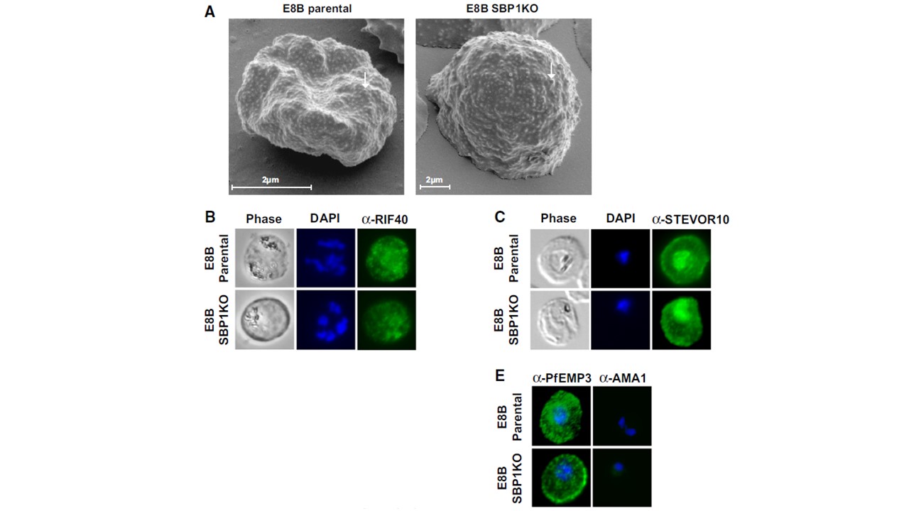
Ultrastructural features of the IE membrane and expression of IE membrane proteins by E8B parasites. a Scanning electron microscopy confirmed the expression of surface knob protrusions (arrows) with IEs from E8B SBP1KO similar to that of E8B parental. Representative images are shown and scale bar represents 2 mm in all images. Labeling of RIFIN (b) and STEVOR (c) proteins expressed by pigmented trophozoite-IEs were visualized by immunofluorescence microscopy using specific a-RIF40 and a-STEVOR10 antibodies (green). Despite the lack of PfEMP1 expression, RIFIN and STEVOR were detected in E8B SBP1KO (lower panel), similar to E8B parental (upper panel). The pattern of staining was consistent with the reported labeling of RIFIN and STEVOR in the IE membrane. e Pigmented trophozoite-IEs of E8B parental and E8B SBP1KO were probed with a-PfEMP3 antibodies (first column) as a positive control and a-AMA1 antibodies (second column) as a negative control (antibody staining in green, DAPI staining in blue). As expected, the pattern of staining was consistent with the labeling of PfEMP3 in the IE membrane and there was no apparent labeling by anti-AMA1. In all immunofluorescence assays, cells were fixed with a mixture of acetone (90 %) and methanol (10 %) and DAPI was used to stain nuclear DNA (blue). All images were taken at equal exposure for both parasite lines and representative images are shown.
Chan JA, Howell KB, Langer C, Maier AG, Hasang W, Rogerson SJ, Petter M, Chesson J, Stanisic DI, Duffy MF, Cooke BM, Siba PM, Mueller I, Bull PC, Marsh K, Fowkes FJ, Beeson JG. A single point in protein trafficking by Plasmodium falciparum determines the expression of major antigens on the surface of infected erythrocytes targeted by human antibodies. Cell Mol Life Sci. 2016 Nov;73(21):4141-58.
Other associated proteins
| PFID | Formal Annotation |
|---|---|
| PF3D7_1133400 | apical membrane antigen 1 |
| STEVOR | STEVOR |
| RIFIN | RIFIN |
