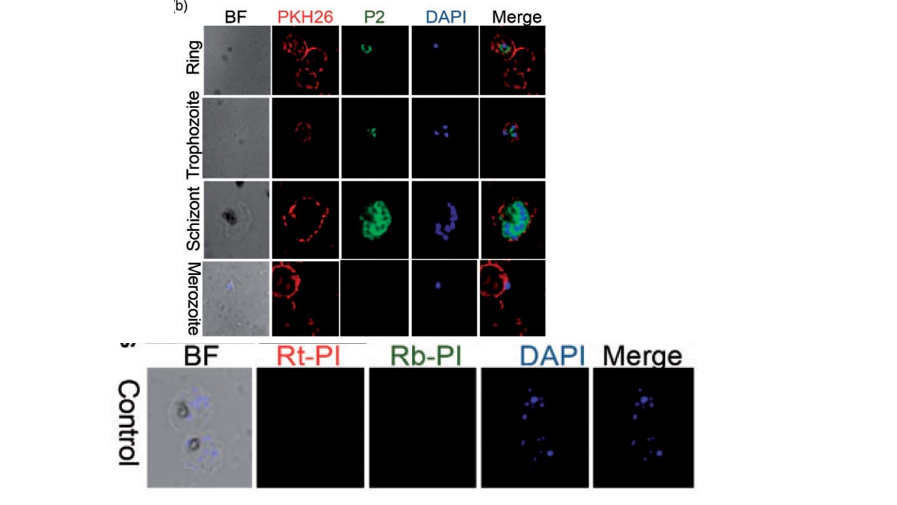
PF3D7_1459400 protein expression profile in asexual stages (b). Stage-specific IFAs performed under permeabilized conditions show that a-PF3D7_1459400 P2 rabbit antibody (green) (1:50) labels rings, trophozoites, and schizonts, but no visible fluorescent signal was observed during merozoite attachment to human erythrocytes. PKH26, the red fluorescent cell linker dye was used to demarcate the erythrocyte membranes. (c). Dual immunofluorescence labeling using a-PF3D7_1459400 P2 rabbit antibody (green), (1:50) and the marker antibody, anti-Plasmodium falciparum Acylated Pleckstrin Homology domain-containing protein (a-PfAPH) rat antibody (red), (1:50) revealed that PF3D7_1459400 protein could be localized on microneme surface. Both antibodies labeled ring, trophozoite, schizont, and released merozoites. Rabbit and rat pre-bleed sera were used as negative controls and no detectable fluorescent signals were observed. The secondary antibody used is Alexa 488-conjugated goat a-rabbit IgG, (1:100; Life technologies, Eugene, USA). Alexa 568-conjugated goat a-rat IgG (red), (1:100); Life technologies, Eugene, USA). Nuclei were stained with DAPI (blue). Exposure times were identical for all images of the same channel. BF: bright field; DAPI: 4,6-diamidino-2-phenylindole); Rt-PI: rat pre-immune; Rb-PI: rabbit preimmune;
Amlabu E, Nyarko PB, Opoku G, Ibrahim-Dey D, Ilani P, Mensah-Brown H, Akporh GA, Akuh OA, Ayugane EA, Amoh-Boateng D, Kusi KA, Awandare GA. Localization and function of a |Plasmodium falciparum protein (PF3D7_1459400) during erythrocyte invasion. Exp Biol Med (Maywood). 2020 5:1535370220961764.
Other associated proteins
| PFID | Formal Annotation |
|---|---|
| PF3D7_1459400 | conserved Plasmodium protein, unknown function |
