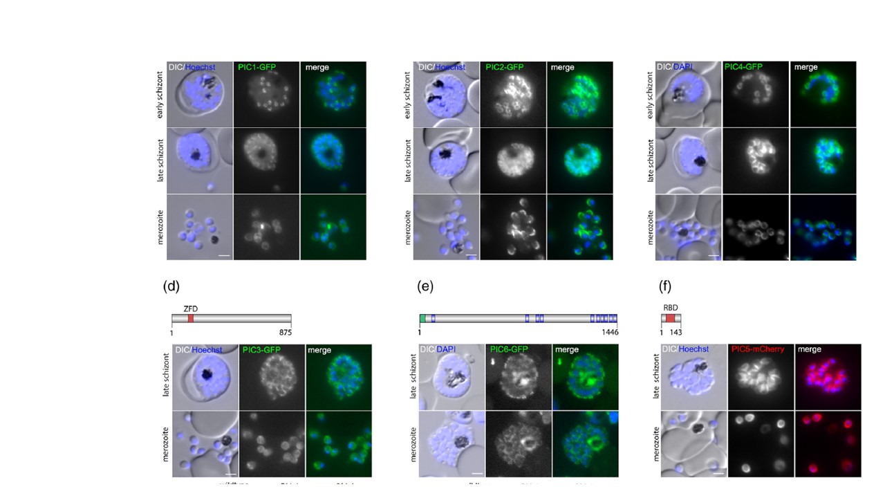
Localization of PhIL1 interacting candidates (PIC) using endogenous GFP tagging. (a) PIC1 (PF3D7_1229300), (b) PIC2 (PF3D7_0822900), (c) PIC4 (PF3D7_0308300), (d) PIC3 (PF3D7_0415800), (e): PIC6 (PF3D7_0530300): On top a schematic representation of the protein (protein length indicated as number of amino acids) with putative protein domains (blue, transmembrane domain; green, signal peptide; red, Zinc Finger domain (ZFD) or RBD (RNA binding domain). Localization of PIC-GFP fusion proteins in schizonts and free merozoites are shown in the middle panel, the PCR-based confirmations of the correct insertion of GFP-encoding integration plasmid into the targeted loci on the bottom. Ladder size indicated in base pairs (bp). gDNA from parental 3D7 was used as control. (f) Localization of PIC5-mCherry (PF3D7_1310700) fusion proteins episomally expressed under the control of the late-stage promoter ama1 in schizonts and free merozoites. Nuclei stained with Hoechst-33,342 or DAPI. Zoom factor: 400%. Scale bar 2 μm. KI, knock-in
Other associated proteins
| PFID | Formal Annotation |
|---|---|
| PF3D7_0109000 | photosensitized INA-labeled protein PHIL1 |
| PF3D7_0308300 | phil1-interacting candidate pic4 |
| PF3D7_0530300 | phil1-interacting candidate pic6 |
| PF3D7_0822900 | phil1-interacting candidate pic2 |
| PF3D7_1229300 | phil1-interacting candidate pic1 |
