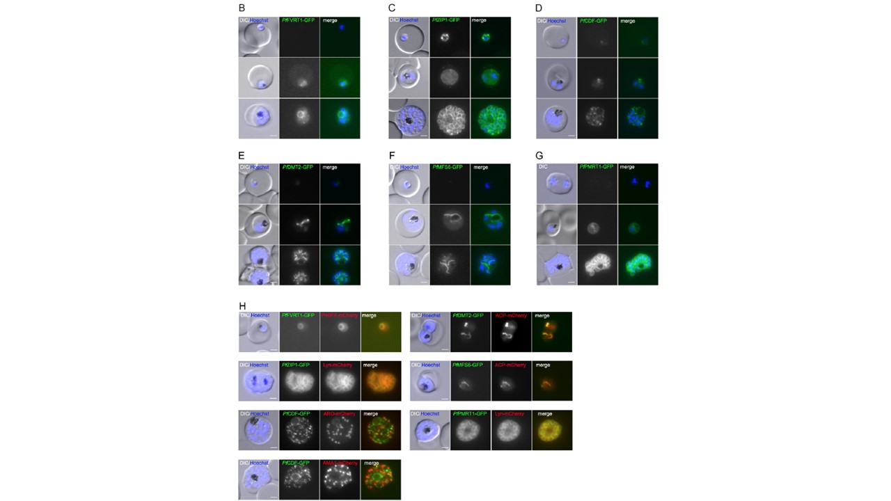
Subcellular localization of six putative P. falciparum transporters during asexual blood stage development. (A) Schematic representation of endogenous tagging strategy using the selection-linked integration system (SLI). Pink, human dihydrofolate dehydrogenase (hDHFR); gray, homology region (HR); green, green fluorescent protein (GFP) tag; dark gray, T2A skip peptide; blue, neomycin resistance cassette; orange, glmS cassette. Stars indicate stop codons, and arrows depict primers (P1 to P4) used for the integration check PCR. (B to G) Localization of (B) PfFVRT1-GFP-glmS, (C) PfZIP1-GFP-glmS, (D) PfCDF-GFP-glmS, (E) PfDMT2-GFP-glmS, (F) PfMFS6-GFP-glmS, and (G) PfPMRT1-GFP-glmS by live cell microscopy in ring, trophozoite, and schizont stage parasites. Nuclei were stained with Hoechst 33342. (H) Colocalization of the GFP-tagged putative transporters with marker proteins P40PX-mCherry (food vacuole), ACP-mCherry (apicoplast), Lyn-mCherry (parasite plasma membrane), ARO-mCherry (rhoptry), and AMA1-mCherry (microneme) as indicated. Nuclei were stained with Hoechst 33342. Scale bar, 2 μm.
Other associated proteins
| PFID | Formal Annotation |
|---|---|
| PF3D7_0609100 | zinc transporter zip1, putative |
