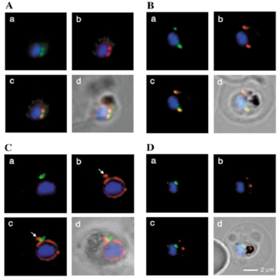
Fluorescence microscopy on fixed parasites. (A) PfGRASP-GFP colocalises with antiPfGRASP-specific antibodies. PfGRASP-GFP is tightly confined to two compartments (a, green) near the parasite nucleus (a, blue). AntiPfGRASP-specific antibodies show a similar staining pattern (b, red with nucleus in blue). Merged image shows identical colocalisation. (B) PfGRASP-GFP colocalises with the cis-Golgi marker ERD2. PfGRASP-GFP (a, green) accumulates in two discrete compartments in close proximity to the nucleus (a, blue). Anti-PfERD2 antibodies recognize similar structures (b, red with nucleus in blue). Merged image shows colocalisation of compartments (c, yellow). (C) PfGRASP-GFP defines a compartment that is distinct from the ER. At the early stages of the parasite life cycle (<16 hours post invasion) PfGRASP is restricted to one compartment (a, green) juxtapose to the nucleus (a, blue). The ER is visualised by anti-PfBiP-specific antibodies (b, red). The membranous system of the ER forms an envelope around the nucleus (b, blue) with one protrusion (indicated by arrow). Merged image shows no colocalisation of the two compartments (c). (D) PfGRASP-GFP does not colocalise with the trans-Golgi marker PfRab6. PfGRASP accumulates in two discrete foci (a, green) adjacent to the nucleus (a, blue). Antibodies against PfRab6 visualise two distinct sites within the parasite (b, red with nucleus in blue). Merged image shows no colocalisation of the PfGRASP defined compartment with PfRab6 (c).
Struck NS, de Souza Dias S, Langer C, Marti M, Pearce JA, Cowman AF, Gilberger TW. Re-defining the Golgi complex in Plasmodium falciparum using the novel Golgi marker PfGRASP. J Cell Sci. 2005 118(Pt 23):5603-13.
Other associated proteins
| PFID | Formal Annotation |
|---|---|
| PF3D7_1017300 | golgi re-assembly stacking protein 2 |
| PF3D7_1144900 | ras-related protein Rab-6 |
| PF3D7_1353600 | ER lumen protein retaining receptor |
