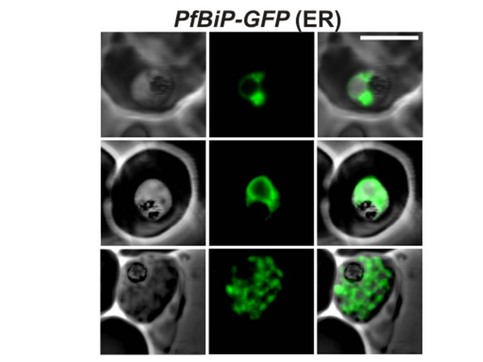
Confocal fluorescence microscopy images of transfected 3D7 P. falciparum-infected RBCs expressing a GFP chimera of PfBiP (GRP78) directed to the endoplasmic reticulum. DIC image, the GFP fluorescence signal and an overlay of a P. falciparum immunoglobin binding protein-GFP. Scale bar = 5 µm. In the ring and trophozoite stages (top and middle rows), the endoplasmic reticulum appears as a ring around the nucleus. In schizont stage parasites fluorescence is observed around the nucleus of individual merozoites (daughter cells).
van Dooren GG, Marti M, Tonkin CJ, Stimmler LM, Cowman AF, McFadden GI. Development of the endoplasmic reticulum, mitochondrion and apicoplast during the asexual life cycle of Plasmodium falciparum. Mol Microbiol. 2005 57:405-19
