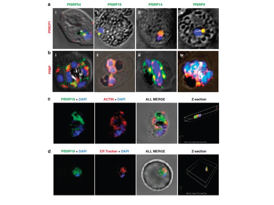
Co-staining of PfSRP polypeptides with nucleolar marker PfNOP1, ER marker PfBiP and cytoplasmic marker Pfactin. (a, b and c) P. falciparum infected erythrocytes stained with anti-PfSRP antibodies (green), anti-PfNOP1 (red), anti-PfBiP (red) and anti-Pfactin (red) respectively. (d) localization of GFP fused SRP19 (green) with ER tracker (red). DAPI is used for nuclear staining (blue). A considerable overlap in staining was observed between three PfSRP polypeptides; PfSRP19, -14, and -9 with PfNOP1 and with PfBiP protein (a and b, i-iv). Importantly, considerable co-localization of PfSRP54 with PfNOP1 in early stages of parasite development i.e. merozoite and ring stages.
Panchal M, Rawat K, Kumar G, Kibria KM, Singh S, Kalamuddin M, Mohmmed A, Malhotra P, Tuteja R. Plasmodium falciparum signal recognition particle components and anti-parasitic effect of ivermectin in blocking nucleo-cytoplasmic shuttling of SRP. Cell Death Dis. 2014 Jan 16;5:e994.
Other associated proteins
| PFID | Formal Annotation |
|---|---|
| PF3D7_0729000 | signal recognition particle subunit SRP9 |
| PF3D7_1203200 | signal recognition particle subunit SRP14 |
| PF3D7_1216300 | signal recognition particle subunit SRP19 |
| PF3D7_1246200 | actin I |
| PF3D7_1450100 | signal recognition particle subunit SRP54 |
