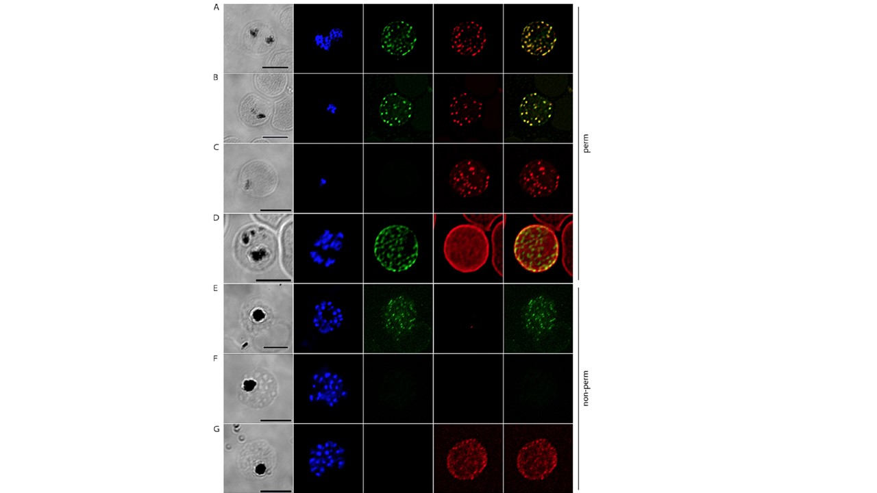
Erythrocyte surface localization of PfEMMA1 by immunofluorescence confocal microscopy. (A–E) Representative Pf3D7-infected RBCs were probed with mouse anti-PfEMMA1 fragment 2 (green) and rabbit antibodies against various parasite or host proteins (red) and counterstained with DAPI to label parasite nuclei. PfEMMA1 colocalizes with REX1 (A) and SBP1 (B), structural proteins ofMC, in permeabilized iRBCs. A concurrent control experiment with mouse preimmune sera is shown (C). PfEMMA1 colocalizes with GPA, a host cell surface glycoprotein, in permeabilized iRBCs (D). In live, nonpermeabilized iRBCs, anti-PfEMMA1 fragment 2 antibodies specifically labeled proteins on the exofacial surface of 8.5% of ~2,000 RBCs (E); rabbit anti-PfMSP4 antibodies did not penetrate the RBC surface membrane. (F and G) Concurrent control experiments with mouse preimmune sera are shown counterstained with anti-PfMSP4 antibodies, which do not penetrate the RBC surface membrane (F), and anti-GPC, which labels exofacial surface glycoproteins (G). Scale bar, 5 μm. DIC,
differential interference contrast microscopy; perm, permeabilized; nonperm, nonpermeabilized; PfMSP4, merozoite surface protein 4; PfREX1, ring exported
protein 1; PfSBP1, skeleton binding protein 1.
Michelow IC, Park S, Tsai SW, Rayta B, Pasaje CFA, Nelson S, Early AM, Frosch AP, Ayodo G, Raj DK, Nixon CE, Nixon CP, Pond-Tor S, Friedman JF, Fried M, Duffy PE, Le Roch KG, Niles JC, Kurtis JD. A newly characterized malaria antigen on erythrocyte and merozoite surfaces induces parasite inhibitory antibodies. J Exp Med. 2021 218(9):e20200170.
Other associated proteins
| PFID | Formal Annotation |
|---|---|
| PF3D7_0207000 | merozoite surface protein 4 |
| PF3D7_0501300 | skeleton-binding protein 1 |
| PF3D7_1134300 | erythrocyte membrane and merozoite antigen emma1 |