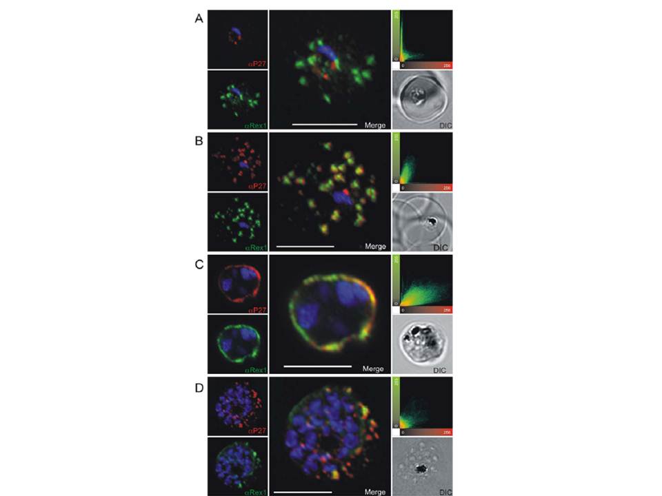
Co-localization of Tex1 with Rex1. P27-specific polyclonal mouse sera (in red) was used to detect TEX1. Rex1 polyclonal rabbit sera (in green). (A) Ring stage parasites; (B) trophozoite stages; (C) schizont stages. Scatter plots show the degree of co-localization of the Tex1 with Rex1 signal. Nuclear DNA was stained with DAPI (blue), Transmission image (DIC), Scale bar: 5 mm. Based on the co-localization of Tex1 and Rex1, Tex1 is localized to the Maurer’s clefts.
Kulangara C, Luedin S, Dietz O, Rusch S, Frank G, Mueller D, Moser M, Kajava AV, Corradin G, Beck HP, Felger I. Cell biological characterization of the malaria vaccine candidate trophozoite exported protein 1. PLoS One. 2012;7(10):e46112.
PubMed Article: Cell biological characterization of the malaria vaccine candidate trophozoite exported protein 1
Other associated proteins
| PFID | Formal Annotation |
|---|---|
| PF3D7_0603400 | trophozoite exported protein 1 |